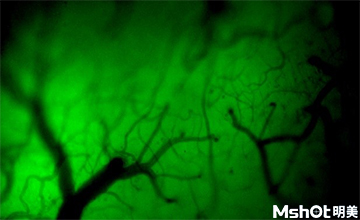
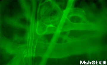
Small animal in vivo fluorescence imaging technology is widely used at home and abroad. More and more researchers hope to track and observe the growth of tumor cells in vivo and the response to drug therapy for a long time through this technology, and hope to observe the distribution and metabolism of fluorescent labeled peptides, antibodies and small molecule drugs in vivo.
Recently, the research-level fluorescence microscope MF43-N independently developed by Mingmei, together with the microscope camera MS23, came into China Medical University to observe the cerebral arteriovenous fluorescence staining of living mice. It was excited by LED fluorescence light source B, and applied to the fluorescence scintillation and the blood cell flow of arteriovenous after the brain staining of living mice. The imaging effect is remarkable. The infinity achromatic independent correction optical system ensures the sharpness, clarity and color restoration of the collected image, which meets the observation needs of researchers. The independent research and development ability has been highly recognized.

