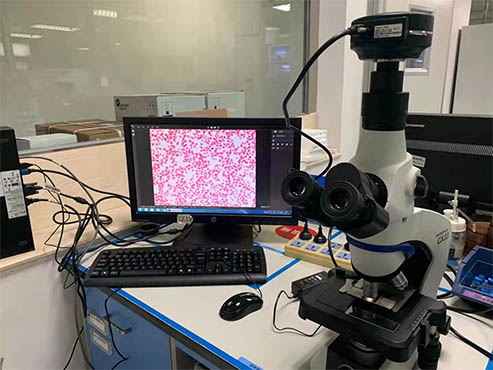
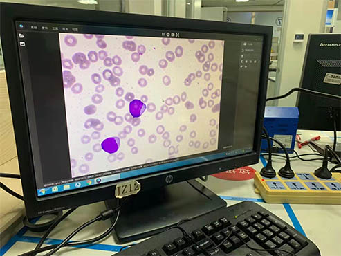
Blood cells are cells that exist in the blood and can flow throughout the body with the flow of blood. This time, Dongguan People's Hospital used an Olympus biological microscope to observe abnormal blood cells. They needed a microscope camera to take a picture of the cell and save it to the computer for comparative analysis and research.

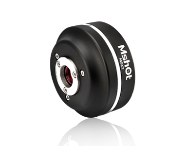
In the end, the staff of Dongguan People's Hospital chose the MSHOT scientific-grade microscope camera MSX2, which can clearly take pictures of abnormal blood cells and make judgments.

The microscope camera MSX2 uses a large-target high-performance imaging sensor, and a USB3.0 data transmission interface is designed. It has the characteristics of high resolution, accurate color reproduction and high sensitivity. It is Ideal tool for liquid-based cell analysis, immunohistochemistry, bone marrow cell analysis, etc. pathological diagnosis with high color requirements.In addition, it also performs in bright and dark fields, phase contrast, polarized light, DIC, fluorescence imaging and other fields.

Micro-shot, since its establishment 19 years ago, has been insisting on honest management and dedicated service, so that the MSHOT brand has formed a good reputation among domestic and foreign universities, research institutes, medical and corporate customers.
If you are interested or have questions about microscope cameras, welcome to contact us!