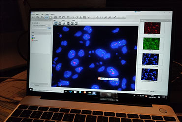
Fluorescence in situ hybridization technology, also known as FISH technology (hereinafter referred to as FISH). FISH technology uses the complementarity of DNA base pairs to hybridize the directly labeled fluorescent single-stranded DNA (probe) with the DNA of the target sample (sample on glass slide) that is complementary to it, and reflects the position of the fluorescent signal on the chromosome by observing the corresponding chromosomes. FISH is a research method that uses this principle to hybridize labeled probes with tissue, nucleus or chromosomal DNA, and conduct qualitative, localization or relative quantitative analysis of the nucleic acid to be detected through a fluorescent detection system.
This technique has the advantages of simple operation, high sensitivity and fast detection speed. The stability of the probes is better than that of radiolabeled probes. At present, it is widely used in Tumor biology, cytogenetics, DNA localization, chromosomal structural variation research and other fields.

It is a common gynecological malignancy, and its incidence ranks high among female malignancies and is on the rise. For the prevention of this malignant tumor, the first principle is "early detection can lead to early diagnosis and treatment". Therefore, in the process of malignant tumor development, the detection of cytogenetic changes is an important means of early detection and early diagnosis and treatment of malignant tumors.
It is a combination of cell in situ molecular hybridization and fluorescence technology. FISH technology can be used to conduct cytogenetic research on abnormal chromosome structure, abnormal chromosome number and DNA localization in solid tumors. FISH technology links cytogenetically altered cell types, locations, and histological structures, and can be used to study the relationship between genetic material and the dynamic changes of malignant tumors, which is helpful for early detection of cervical cancer tumor cells and timely diagnosis and treatment.

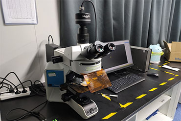
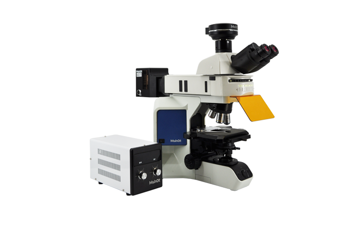
Research-grade upright fluorescence microscope MF43-N, is equipped with a high-power four-channel LED fluorescent light source, which has the advantages of less heat generation, high power, strong light source brightness, long life and easy maintenance. In addition, the turntable fluorescence switching method allows users to flexibly switch fluorescence. Users can also choose to use our high-sensitivity microscope camera, which can quickly help users to capture fluorescent signals. Combined with MSHOT's FISH software, users can get detection results in a very short time.

If you would like to get more information, you could visit www.m-shot.com.