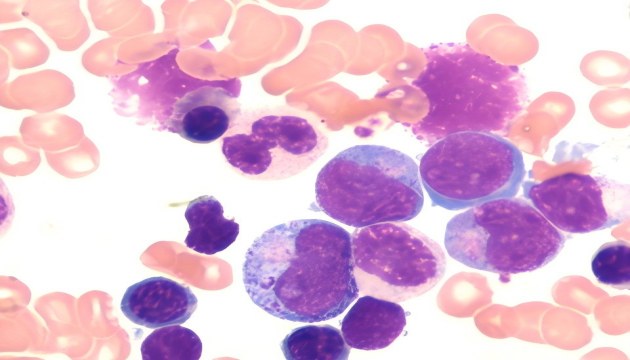
A blood smear is the fundamental method for hematocytological examination. By observing the morphology of peripheral blood cells under a microscope, it screens for abnormal blood cells and pathogens such as bacteria and parasites, assisting in the diagnosis of diseases including anemia, infections, and leukemia.
Microimaging is used in blood smears to clearly observe the morphological details of blood cells such as red blood cells, white blood cells and platelets. Staining techniques are applied to enhance contrast, helping to identify abnormal cells or pathogens and improve diagnostic accuracy.
Challenges of Microimaging in Blood Smears
Challenges of microimaging in blood smears include distinguishing cells with similar morphologies (such as platelets and malaria parasites), as well as maintaining image clarity and color reproduction under high-power magnification to ensure accurate diagnosis.
Solutions for Microimaging in Blood Smears
● High-Quality Observation Solution: Recommended ML51-N equipped with MSX2/MSX11 cameras, semi-apo chromatic objectives. It delivers clear and detailed high-power imaging, and captures fine details with high-pixel imaging, improving diagnostic efficiency and accuracy.
● Cost-Effective Solution: Recommended ML31 equipped with MDX10/MSX2 cameras. Adopting plan achromatic objectives and LED Köhler illumination, it provides images with excellent clarity and accurate color reproduction.