
Pathology Imaging
Modern medical diagnostic methods are highly diverse. Commonly used approaches range from analytical diagnoses based on medical history, symptoms, and clinical signs to imaging-based diagnoses using ultrasound, CT, MRI, and other modalities. However, a definitive diagnosis often requires confirmation by the pathology department through pathology imaging.
Pathological diagnosis, which is made by observing gross (macroscopic) changes in organs and examining tissue structures and cellular pathological features under a microscope, is more objective and accurate. An objective and accurate pathological diagnosis relies not only on the professional judgment of pathologists but also on high-performance optical microscopes to obtain precise and clear pathological images.

Applications of Pathology Imaging
Pathology is classified into organ pathology, tissue pathology, and molecular pathology based on the objects of observation.
Organ pathology relies on gross examination and microscopy to determine the nature of lesions.
Tissue pathology observes subtle lesions through section staining; common methods include HE, PAS, Gram, Papain, and Oil Red O staining.
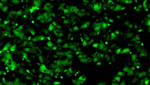
Molecular pathology analyzes protein or gene expression for disease progression and drug efficacy evaluation.
Common techniques involve IHC (Immunohistochemistry) for protein localization and FISH (Fluorescence In Situ Hybridization) for detecting specific gene fragments, supporting precision medicine.
Challenges in Pathology Imaging
Pathology imaging faces challenges in ergonomics, flatness of field, color reproduction, and fluorescence signal acquisition. It requires ergonomic design, flat-field objectives, precise color reproduction technology, and long-life LED light sources.
Ergonomic design improves operational comfort and reduces fatigue during long-term microscopy. Flat-field objectives can achieve a flat field of view with a field number (FN) of 22 mm or even 25 mm, greatly enhancing microscopy efficiency.
Color reproduction depends mainly on the light source and camera. The use of high-color-rendering LED light sources matched with microscopes and cameras that deliver excellent color fidelity helps ensure accurate color reproduction.
In addition, for the scanning of small numbers of digital slides, MSHOT provides a slide scanning system that quickly synthesizes high-definition digital slides, balancing large field of view and fine details to improve analytical efficiency.

Solutions for Pathology Imaging
- Recommended ML31-M/ML51-M microscopes with MSX series cameras: ergonomic design reduces fatigue, while wide-field eyepieces and high-definition objectives improve efficiency.
- For FISH/immunofluorescence observation, the MF43-N fluorescence microscope is equipped with a multi-channel fluorescence turret, long-lasting LED light source, high-resolution objectives, and is compatible with large-pixel cameras.
- MDS4 digital slide scanning system integrates scanning and fluorescence functions, enabling fast stitching of high-definition large images, suitable for scientific research and clinical use.
- ME40 macro camera features auto-focus, high pixels, high zoom, foot pedal control, and flexible installation, ideal for gross specimen documentation.