
In the field of environmental pollutant research, the combined toxicity of nanoplastics (NPs) and persistent organic pollutants has long been a focal point for scientists. Recently, the research team led by Wang Jun from South China Agricultural University published a significant research finding in Journal of Hazardous Materials, a top – tier journal in environmental science (CAS Category 1, IF = 12.2).
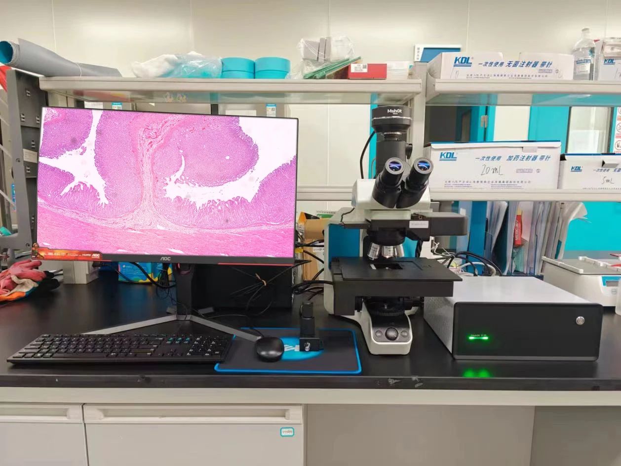
This study reveals the synergistic toxic effects of nanoplastics and 6:2 chlorinated polyfluoroether sulfonate (F53B) in the presence of natural organic matter (NOM). Notably, throughout the research, the digital slide scanning system MDS4 from Mshot Optoelectronics was used to conduct histopathological imaging analysis, providing high – precision visual support for the conclusions!

Mshot MDS4 Helped Accurately Capture Toxicity Evidence
1.High - resolution imaging intuitively presents tissue damage
The study, through a 21 – day chronic exposure experiment, found that in the presence of NOM, NPs and F53B significantly exacerbate liver and intestinal damage in zebrafish. High – definition digital sections taken by Mshot MDS4 (Figure 1) clearly show:
Liver: Vacuolization, nuclear condensation
Intestine: Villus fusion, reduced crypt depth

Figure1:Histopathological changes of liver and intestinal tissues in adult zebrafish after long-term exposure to different treatment groups for 21 days. (A) Schematic diagram of the histological morphology and structure of liver cells and intestinal villi; (B) Control group – C; (C) 1 mg/L NPs – AN; (D) 200 μg/L F53B – AF; (E) 1 mg/L NPs + 200 μg/L F53B – ANF; (F) 1 mg/L NPs + 200 μg/L F53B + 10 mg/L HA + 10 mg/L BSA – M. Among them, VL is villus length, CD is crypt depth, TMT is muscle layer thickness, the black arrow indicates liver cell nuclear shrinkage, and the black frame indicates intestinal villus atrophy.
2.Multi - dimensional Analysis of Toxicity Mechanisms
Oxidative Stres:In the M group (NPs + F53B + NOM), the level of malondialdehyde (MDA) significantly increased, while total antioxidant capacity (T-AOC) decreased.
Gene Regulation:Inflammatory-related genes (e.g., il1b) were upregulated.Immunosuppressive genes (e.g., nfkbiaa) were downregulated.
Intestinal Microbiome Dysregulation:The abundance of pathogenic bacteria (e.g., Clostridium) increased.
3.Transcriptomic Validation
The pathological results assisted by Mshot MDS4 are highly consistent with the transcriptome data, confirming that NOM exacerbates toxicity by activating the AMPK/PPAR signaling pathway.
Why choose Mshot MDS4?
The Powerful Capabilities of Research-Grade Equipment
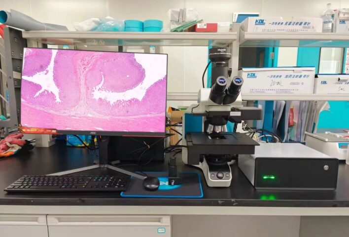
The key pathological data in this study all depend on the Mshot Digital Slide Scanning System MDS4. Here’s why it stood out:
- Dual – camera solution, enabling high – definition single – field imaging & bright – field whole – slide scanning imaging
- Under the 20X objective lens, can clearly identify micron – level lesions such as nuclear shrinkage, villus atrophy, etc.
- Upgradeable for fluorescence observation; integrates bright – field slide scanning and research – grade fluorescence microscopy into one, dual – use with a single instrument
- Scanning speed and precision comparable to box – type scanners
- Professional storage format and slide – viewing software; digital archiving facilitates long – term analysis and result reproduction
- High – precision three – axis motorized control, while retaining manual focusing
- 40s rapid scanning, optional single – slide or 5 – slide scanning
Mshot has always been committed to providing high – end microscopic imaging solutions for life sciences and medical testing. The research achievements of South China Agricultural University this time have once again verified the value of Mshot equipment in the study of complex toxicity mechanisms:
Application scenarios: Environmental toxicology, pathology, microbial ecology
Technology extension: MDS4 is compatible with multi – modal imaging such as fluorescence and bright field, and adapts to the needs of cutting – edge research topics
In the future, we will continue to cooperate with scientific researchers, using precise imaging technology to reveal the mysteries of the microscopic world!