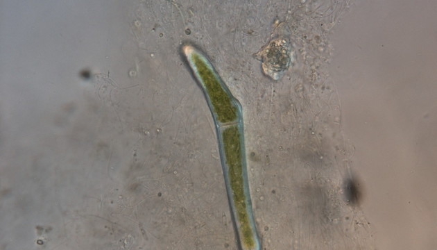
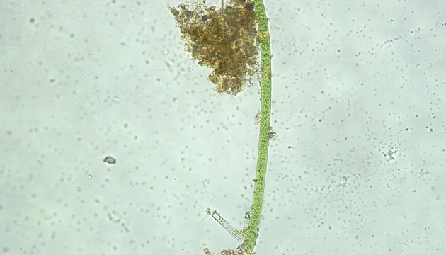
Algae are a group of eukaryotic or prokaryotic organisms (Phylum Cyanophyta, i.e., cyanobacteria) belonging to the Kingdom Protoctista. They are mostly aquatic, lack true roots, stems and leaves, and have no vascular bundles, but are capable of photosynthesis. They vary greatly in size, ranging from unicellular flagellates as small as 1 micrometer in length to large brown algae up to 60 meters long.
Applications of Microscopic Imaging in Algal Research
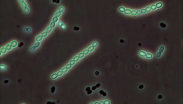
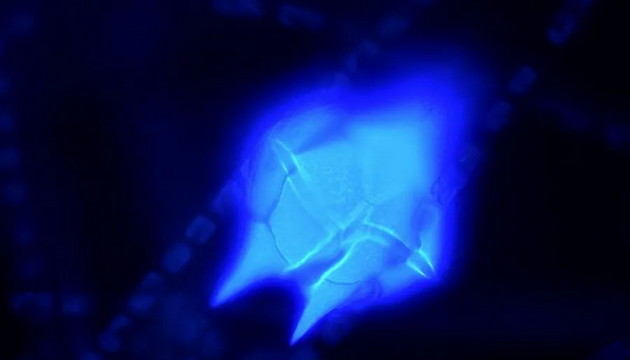
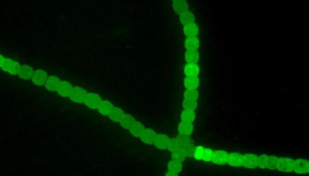
Brightfield microscopy is commonly used for algal monitoring to observe species and quantity. For algae with indistinct pigments, such as diatoms, phase contrast or DIC can be used to enhance contrast. For specific algae such as cyanobacteria and green algae, fluorescence microscopy can be used to observe fluorescence signals under specific light, enabling rapid identification.
Pain Points in Algal Research
Challenges in algal monitoring include: difficulty in identifying tiny algae, the trade-off between imaging depth of field and resolution, and fatigue during long-term observation. High-resolution objectives, depth of field synthesis technology, and ergonomically designed microscopes are required to improve monitoring efficiency and comfort
Solutions for Algal Research
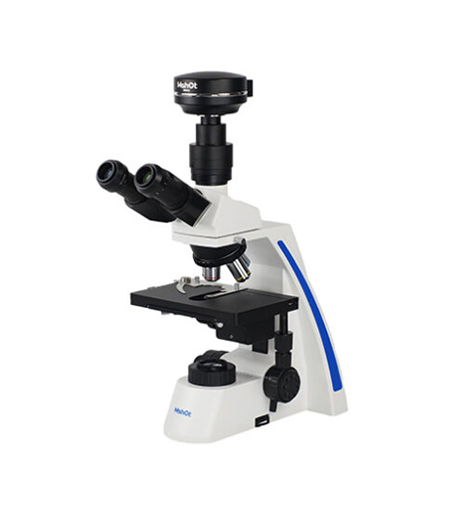
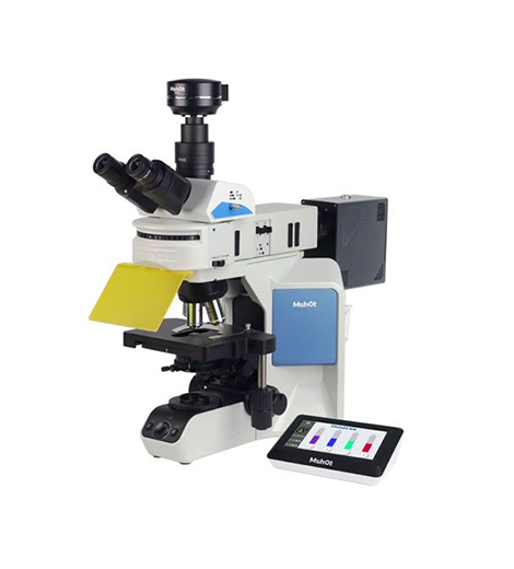
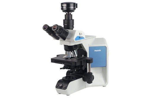
Brightfield Observation Solution: biological microscopes ML31/ML43/ML51-N, equipped with wide-field eyepieces, high-quality plan achromatic objectives/semi-apochromatic objectives, providing excellent brightfield and phase contrast imaging. Fluorescence Observation Solution: Fluorescence microscopes MF31/MF43-N with LED fluorescence excitation, easy to operate. MF43-N supports optional fluorescence excitation blocks, enabling 5-color fluorescence excitation to flexibly meet various monitoring requirements.