Home » Microscopy Applications in Virology Laboratories of CDC
Microscopy Applications in CDC Virology Laboratories
Virus “Scouts” — Unveiling Microscopy Applications in CDC Virology Laboratories
As the global landscape of infectious disease prevention and control grows increasingly complex, the virology laboratories of the Centers for Disease Control and Prevention (CDC) have become ever more vital in public health, playing an irreplaceable and critical role in infectious disease control.
Optical microscopes are widely and deeply applied in CDC virology laboratories. They serve an irreplaceable function in virus detection, mechanistic research, and prevention support, providing robust backing for the advancement of virology and public health preparedness.
Applications of Microscopes in Virology Laboratories
Preliminary screening with an inverted microscope
Daily observation of cell morphological changes and documentation of lesion progression (e.g., vacuolization, cell detachment).
Analysis with a fluorescence microscope
Precise localization of virus-infected regions using fluorescently labeled antibodies or dyes.
Challenges of Microscopic Imaging in Virus Observation
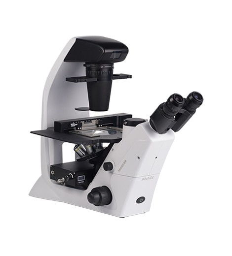
Viruses typically measure less than 100 nm in size, far exceeding the resolution limit of optical microscopes (approximately 200 nm). As a result, it is difficult for optical microscopes to directly and clearly observe viral morphology and structure. In practical applications, routine detection relies on indirect methods: preliminary assessment of infection status is made by observing pathological morphological changes in host cells caused by viral infection (e.g., vacuolization, syncytium formation) or tissue damage characteristics.
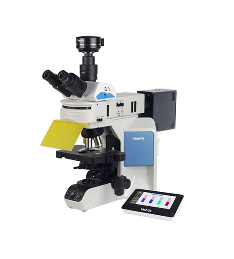
For detection using fluorescence microscopy, specific fluorescent labeling strategies (such as labeling viral antigens with immunofluorescent antibodies or viral genomes with nucleic acid probes) can be employed to localize viral components at the subcellular level. However, due to the tiny volume and low copy number of viral particles, the intensity of the fluorescence signal generated by labeling is significantly limited. To overcome this issue, high-intensity excitation light and microscopes equipped with high-sensitivity cameras are required for imaging to enhance signal capture and recording, thereby improving the accuracy and sensitivity of virus detection.
MSHOT Microscopy Solutions
Respiratory 7-Panel / 9-Panel Testing Solution
Recommended: Upright fluorescence microscopes MF31/MF23 + microscope cameras MD60/MDX10 (primary recommendation). Suitable for fluorescence observation in respiratory 7-panel and 9-panel testing, with B-excitation fluorescence (460–490 nm) displaying green fluorescence reactions.


