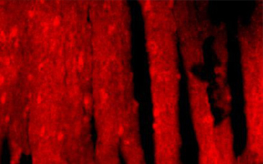
Fluorescence in situ hybridization (FISH) is a non radioactive molecular cytogenetic technology developed on the basis of radioisotope in situ hybridization in the late 1980s. It is a new in situ hybridization method, which is formed by replacing isotope labeling with fluorescent labeling. Fish has the advantages of safety, rapidity, high sensitivity, long-term preservation and multi color display. It can not only display the midphase mitotic phase, but also display the interphase nucleus.
Principle of FISH application
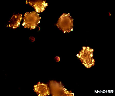
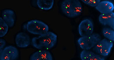
In order to detect the chromosome or gene abnormality in cell or tissue samples, the fluorescence labeled DNA probe in vitro is used to hybridize the probe with the DNA base pair of the sample, and the fluorescence signal is detected by fluorescence microscope.
Technical characteristics of FISH
The operation is simple, the detection is fast, the results can be obtained within 24 hours, and the results are easy to observe.
Good repeatability and accurate spatial positioning.
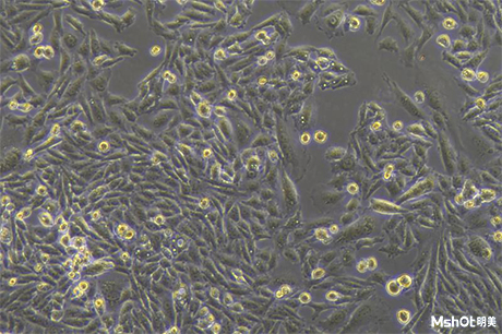
There are abundant sources of specimens: interphase cells, metaphase cells, differentiated or undifferentiated cells and dead or alive cells can be detected.
Personnel training is fast.
Application and clinical significance of FISH application
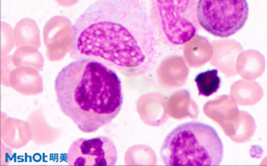
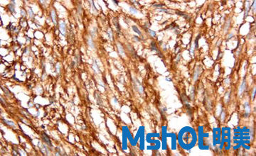
Applications: tumor diagnosis, chromosome research, gene localization, and the combination of immune, quantum dots, microfluidic chips and other technologies.
Clinical significance: guiding medication, early diagnosis, differential diagnosis and evaluating prognosis.
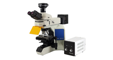
Since MSHOT launched the FISH detection fluorescence microscope and FISH analysis software system in 2011, it has provided fish overall solutions for hundreds of medical institutions in China. During this period, MSHOT fluorescence microscope and FISH analysis software system have been continuously developed and upgraded to better meet the needs of user.
MSHOT FISH solution includes the following aspects
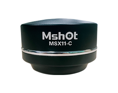
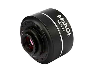
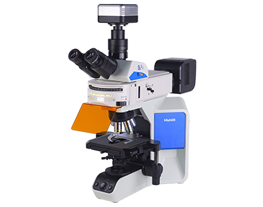
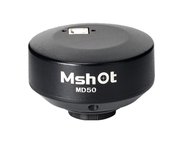
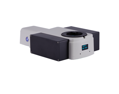
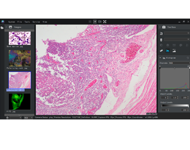
If the user does not have fluorescence microscope and FISH analysis software system, the whole fish fluorescence microscope MF43 / MF43-N and FISH analysis software system MC50-S / MS23-FISH are provided.
When the user has fish fluorescence microscope, it mainly provides FISH analysis software system MC50-S / MS3-FISH.
If the user has imported brand microscope (no fluorescence observation), it will provide fish fluorescence upgrade and FISH analysis software system MC50-S / MS23-FISH.
Provide fluorescent excitation block (including dual pass and multi pass), matching Olympus / Leica / Nikon / Zeiss four brands, suitable for Abbott, IBP(Anbiping), FastProbe (HealthCare), Diaglogic, Celnovte and other FISH probes.
MSHOT user lists and cases (continuously updated)
1. Henan Pingdingshan pingmedia Group General Hospital
2. Henan children's Hospital
3. The second people's Hospital of Jiaozuo City, Henan Province
4. Peking University Shenzhen Hospital
5. Zhengzhou Third People's Hospital
6. Sichuan Guangyuan Central Hospital
7. The Second Affiliated Hospital of Hunan Nanhua University
8. Guangzhou Maijing Gene Technology Co., Ltd
9. Shandong Weihai Central Hospital
10. Henan Sanmenxia Central Hospital
11. Shandong Lanling people's Hospital
12. Affiliated Hospital of Sichuan Panzhihua University
13. Medical College of Wuhan University
14. Xiamen Hongai hospital
15. The First Affiliated Hospital of Lanzhou University
16. Wuxi Second People's Hospital
17. Shanxi Yuncheng First Hospital
18. Luoyang Central Hospital
19. Huaihua Second People's Hospital
20. Wuxi maternal and Child Health Hospital