
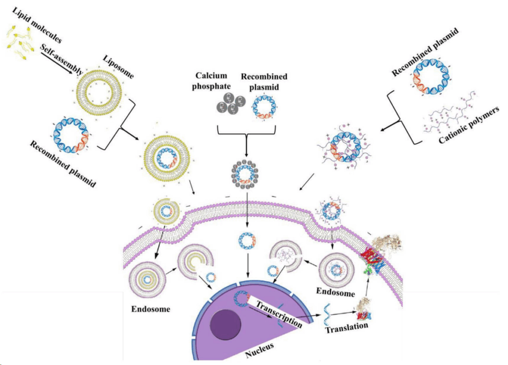
In the journey of exploring the mysteries of gene function, developing new therapies, or producing biological agents, cell transfection technology is like a magical key in the hands of researchers. It accurately delivers exogenous nucleic acids (such as DNA, RNA, and siRNA) into cells, enabling gene manipulation—including overexpression, silencing, and editing.
However, whether this key can successfully unlock the target door often hinges on the accurate evaluation and optimization of transfection efficiency. Traditional detection methods are like “viewing flowers through a fog” (a Chinese idiom meaning to see things vaguely), while the emerging live-cell imaging technology is bringing about a disruptive transformation.
1.Foundations and bottleneck:three limitations
The level of transfection efficiency directly determines the reliability and reproducibility of experimental results.
For decades, researchers have relied on several classic methods for evaluation—each with its own strengths, yet each also facing insurmountable gaps:
Fluorescence Microscopy Observation (Qualitative/Semi-Quantitative)
Advantages:
- Intuitive visualization: Enables direct, clear visual assessment of transfected cells.
- Subcellular localization capability: Allows precise observation of the subcellular distribution of target nucleic acids or proteins.
- Broad equipment accessibility: Fluorescence microscopes are widely available in most laboratories, ensuring easy implementation.
Limitations:
- Static single-time-point analysis: Only provides a “snapshot” of the system at a specific moment, unable to capture the dynamic changes in transfection efficiency over time.
- High subjectivity in analysis: Manual counting of positive cells introduces significant variability across different fields of view, leading to poor reproducibility and difficulty in achieving precise quantification.
- Low throughput: Manual inspection of samples well-by-well is time- and labor-intensive, making it impractical for large-scale screening experiments.
- Phototoxicity risks: Frequent or high-intensity excitation light exposure can damage cells, disrupt their normal physiological states, and ultimately lead to unreliable experimental data.
Flow Cytometry (Quantitative)
Advantages:
- High-throughput, objective, and quantitative analysis: Enables high-throughput, objective, and quantitative analysis of a large number of single cells, providing accurate percentages of transfection efficiency.
Limitations:
- Destructive endpoint assay: Cells need to be trypsinized into a suspension, which terminates subsequent experiments.
- Single-time-point detection: Only provides efficiency data at the moment of detection, failing to track dynamic changes over time.
- Loss of spatial information: Critical spatial information—such as cells’ location in the culture dish, morphological features, and intercellular interactions—is completely lost.
- Detrimental to adherent cells: Causes severe damage to sensitive adherent cells (e.g., primary cells, neurons).
qPCR (mRNA-Level Quantification)
Advantages:
- High sensitivity: Capable of detecting low-abundance mRNA expression (enabling accurate quantification of rarely expressed transcripts).
Limitations:
- Sample destructiveness: Cells are lysed (broken down) during sample preparation, making them unreusable for subsequent experiments.
- Disconnect from protein function: mRNA expression levels do not equate to the expression of active proteins (mRNA abundance does not directly reflect actual protein activity or concentration).
- Lengthy operational workflow: Involves a multi-step process—RNA extraction → reverse transcription → amplification—with errors accumulating at each step.

2.Common Dilemmas of Traditional Methods
“Black Box”-Style Experiments
Changes in cell status inside the incubator are invisible to researchers, who can only passively wait for preset time points to perform measurements—unable to monitor dynamic shifts in real time, much like guessing the contents of an unopened box.
High Optimization Costs
Optimizing transfection conditions (e.g., DNA concentration, reagent ratio, incubation time) requires setting up numerous parallel samples and conducting destructive assays at multiple time points, leading to substantial consumption of labor, materials, and time.
Fragmented Data
Assessments of transfection efficiency, cell toxicity, and functional phenotypes demand separate experiments, resulting in weak correlations between different datasets.
Questionable Physiological Relevance
Cell fixation, trypsinization, and staining severely disrupt normal cellular states, making it challenging for the generated data to fully represent genuine physiological conditions.
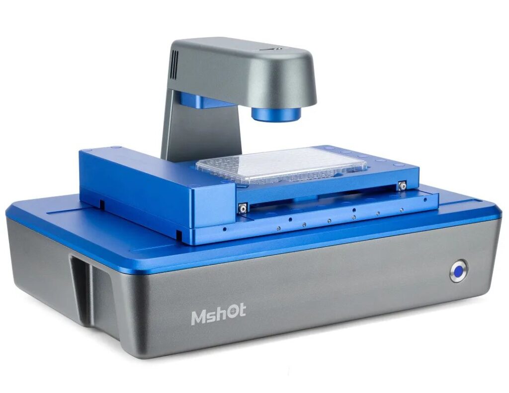
3.Breaking the Deadlock: Four Disruptive Breakthroughs of Mshot MCS31
Faced with the limitations of traditional methods, the Mshot Live Cell Imaging System MCS31 has emerged as a solution. By deeply integrating dynamic imaging, high-throughput scanning, and AI analysis, the Mshot Live Cell Imaging System MCS31 opens up an entirely new dimension for transfection efficiency evaluation, enabling a leap from “static endpoint blind testing” to “dynamic full-process visual intelligent analysis.”
Breakthrough 1:
Real-Time Dynamic Tracking, Ending “Single-Point Blind Testing”
Non-Destructive Observation in Incubators for Extended Periods: Enables continuous, label-free, and low-phototoxicity observation, recording dynamic curves of fluorescent expression.
Key Values:
- Accurately capture the optimal expression time window to avoid “time-point misjudgment.”
- Distinguish between transient transfection (short-lived peak) and stable transfection (sustained high expression).
- Reveal intercellular heterogeneity (observe differences in transfection efficiency among individual cells and their dynamic changes).
Breakthrough 2:
High-Throughput Automation, Achieving Leapfrog Efficiency Improvement
Full-Automated, Rapid Panoramic Scanning for 6–384 Well Plates: Automatically analyzes multiple samples in a single run.
Key Values:
- Revolutionizing Condition Optimization: Test dozens of different transfection condition combinations (e.g., DNA concentration gradients, transfection reagent ratio gradients, time gradients) simultaneously on a single multi-well plate (e.g., 96-well plate). The MCS31 can quickly complete full-plate scanning and automatically analyze the efficiency of each well, significantly accelerating the optimization process.
- Drug/Reagent Screening: Efficiently evaluate the impact of different transfection reagents, auxiliary enhancers, or drugs on transfection efficiency.
Breakthrough 3: Flexible Compatibility and Stable Environment
Compatibility with Multiple Culture Vessels: Supports culture dishes, 6–384 well plates, T-flasks, glass slides, and more.
Full Sealed Design: Ensures long-term operation in a stable cell culture environment with 37°C temperature, 5% CO₂, and saturated humidity, preventing fluorescence signal drift caused by temperature fluctuations.
Key Value:
Enables more flexible experimental design and generates more stable and reliable data.

Breakthrough 4: Ultra-Low Damage, Ensuring Physiological Authenticity
625nm Red Light Technology: Minimal phototoxicity with high cell viability.
Label-Free Phase-Contrast Imaging: Enables observation of cell morphology and migration without staining, resulting in data that more closely reflects the true physiological state.
Key Value:
Transfected cells can be directly used for downstream functional experiments.
4.Transformative Applications of Live-Cell Imaging
The MCS31 is more than just an instrument—it represents an upgrade in transfection research paradigms, demonstrating tremendous potential in cutting-edge fields:
Gene Therapy: Dynamically monitor viral vector transduction efficiency and long-term expression stability.
CRISPR Editing: Real-time tracking of editing efficiency, clonal growth, and cellular stress responses.
High-Throughput Drug Screening: Simultaneously eliminate cytotoxic compounds during reporter gene screening.
Cell Line Development: Dynamically screen production clones with high expression and stable growth.
Breakthrough in Hard-to-Transfect Cells: Provide non-destructive evaluation protocols for primary cells/stem cells.
Cell transfection stands as a cornerstone experiment in unraveling the mysteries of life, with transfection efficiency forming the load-bearing core of this foundation. While traditional detection methods have played invaluable roles, their limitations—static measurements, destructiveness, low throughput, and singular information output—have become bottlenecks constraining research efficiency and depth.
In the pursuit of more precise, efficient, and life-true scientific inquiry, live-cell imaging technologies represented by the Mshot MCS31 undoubtedly embody the future direction of transfection efficiency analysis, with prospects that are infinitely promising.