
1. What is Chromosome Examination?
Chromosomes are carriers of genetic material and play a crucial role in heredity and development. Human chromosomes consist of two types: 22 pairs of autosomes and 1 pair of sex chromosomes, giving a total of 46 chromosomes in each cell. The normal karyotype is represented as “46, XX (or XY)”.

The left image shows the normal karyotype of a female, while the right image shows that of a male
With advancements in medicine and technology, it has been discovered that certain diseases are linked to abnormalities in chromosome structure or number. Using optical microscopes for karyotype analysis allows for the assessment of potential disease risks. Common chromosomal genetic disorders include:
① Trisomy 21 (Down syndrome), which results in intellectual disability and characteristic facial features, with the karyotype being “47, XX (or XY), +21” (an extra chromosome 21).
② Trisomy 13 (Patau syndrome), marked by intellectual disability, cleft palate, malformed ears, and craniofacial deformities, with the karyotype “47, XX (or XY), +13”.
③ Trisomy 18 (Edwards syndrome), characterized by developmental delays and physical retardation, with the karyotype “47, XX (or XY), +18”.
④ Klinefelter syndrome (47, XXY), where males have an additional X chromosome, leading to infertility and other issues.
⑤ Supermale syndrome (47, XYY), where males possess an additional Y chromosome, typically presenting with tall stature, excitability, and irritability.
Chromosome examination is used to detect anomalies in chromosome number or structure and is widely applied in pre-marriage, prenatal screenings, and diagnostic monitoring of systemic diseases. Traditionally, a biological microscope is used to observe the number and structure of chromosomes, which is a quick and simple method. Recently, fluorescence microscopes have been used for chromosome analysis, employing Fluorescence In Situ Hybridization (FISH) technology to detect and label specific chromosomal regions, offering higher precision but more complex operation.
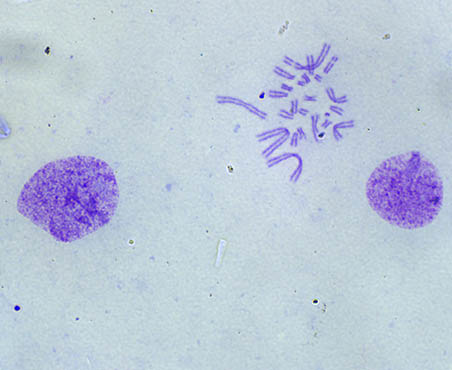
Chromosome samples for examination include peripheral blood, bone marrow cells, skin, and amniotic fluid. Traditional karyotype analysis involves studying chromosomes during metaphase, where the cells are arrested at this stage using colchicine. After a series of cytological treatments such as hypotonic and fixation procedures, the chromosome specimen is stained with dyes like aceto-orcein or gentian violet, then observed under a microscope to determine whether the number or structure of chromosomes is abnormal, helping to assess disease risk.
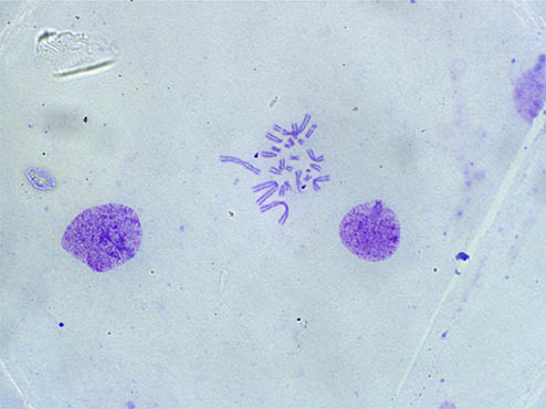
Image of chromosomes taken with the ML51-N microscope paired with MSX11 (100x oil immersion objective)
2. Challenges in Chromosome Microscopy
Human chromosomes typically range in diameter from 0.2 to 2 micrometers, with lengths varying from 0.2 to 50 micrometers. When performing chromosome microscopy with a biological microscope, the primary challenge is distinguishing abnormalities in both the number and structure. The key challenges include the following:
(1) The Small Size of Chromosomes and Its Impact on Resolution
Chromosomes during metaphase are very close to each other, with a centromere connecting each pair. If the objective lens does not have sufficient resolution, it becomes difficult to differentiate between the chromosomes and accurately assess their structure, leading to misinterpretation of their number and structure.

A higher numerical aperture (NA) objective lens translates to a higher resolution. The high NA objectives in Mshot biological microscopes allow for excellent resolution, with the 100x oil immersion objective having a NA of 1.35 and the 40x objective offering a NA of 0.75. These provide theoretical resolutions of 0.3 micrometers and 0.5 micrometers, respectively, making it easy to distinguish the structure and number of chromosomes.
(2) Focus Stability with High Magnification
A 100x objective lens has a small depth of field due to its high resolution. Chromosome samples may be at different focal planes due to variations in preparation, necessitating fine focus adjustments to obtain a clear image. Mshot biological microscopes use coaxial coarse and fine focusing mechanisms with a micro-adjustment step size of 2 micrometers, allowing for precise focusing with minimal risk of over-focusing, making operations smooth and accurate.
(3) Ergonomics for Extended Viewing Periods
Chromosome examination, especially in hospital pathology departments, requires long hours of microscope use. To alleviate fatigue, Mshot research-grade biological microscopes feature ergonomic designs such as a low-position stage, 30° inclined eyepiece, soft and non-glare white LED light source, and a robust, high-rigidity frame. These features reduce eye, hand, and back fatigue during extended periods of observation.
3. Mingmei Solutions
(1) Recommended Biological Microscope for Chromosome Examination: ML31/ML41/ML51-N + MSX11/MSX2

- Ergonomically designed low-position stage and focusing mechanisms to reduce fatigue
- 22mm/25mm wide-field eyepieces offering excellent flat-field performance, ideal for extended observation
- High numerical aperture objectives for superior resolution and reduced imaging blur
- LED brightfield light source with adjustable brightness for comfortable, non-glare illumination
- 21/12 megapixel high-resolution camera paired with professional imaging software for clear details
(2) Recommended Digital Slide Scanning Solution: Mshot Digital Slide Scanning System MDS4

- Suitable for routine brightfield pathology slide scanning such as HE, IHC, TCT
- High-precision 3D motion translation stage with electronic handwheel control
- Fast, automatic autofocus and scanning range recognition
- High frame rate scanning with 10X magnification scans completed in as little as 40 seconds
- Upgradable to include fluorescence functionality, enabling multi-modal slide scanning and fluorescence observation
(3) Recommended Motorized Upgrade: Motorized Stage + Conventional Biological Microscope

- Quantitative selection of regions for karyotype analysis, preventing errors from manual operation
- Effective travel range of 73x30mm with a small step size of 1 micrometer for accurate positioning
- Compact design compatible with most biological microscopes
- Available for secondary development, with a full SDK development package provided
Mshot is a high-tech enterprise specializing in the research, development, and sales of microscopic imaging products. As a member of the Optical Branch of the China Instrument and Meter Industry Association, the company manufactures and sells medical microscopes and related products.
Mshot is committed to honest business practices and providing stable, high-quality products and services for education, research, medicine, and industrial inspection.
With customer needs as a priority, quality as the foundation, and innovation as the direction, Mshot is driving the digital, motorized, and intelligent transformation of the microscopy industry, contributing to the domestic development of the microscope sector.
If you are interested in microscopy imaging products or have any questions, Mshot welcomes your inquiries and looks forward to collaborating with you!