
In cell biology and cancer research, there exists a classic yet often overlooked experiment – the Wound Healing Assay. By artificially creating a “wound” in a confluent cell monolayer, researchers observe how cells migrate, proliferate, and gradually repair the damaged area under different conditions. This reveals crucial insights into cellular migration capacity, repair rates, and even the efficacy of drug interventions.
Application Scenarios and Traditional Methodology

The application scenarios of the wound healing assay are extensive, including but not limited to:
Tumor Cell Metastasis Mechanisms
Determining cancer cell invasiveness and the inhibitory effects of exogenous agents on their migration.
Inflammation and Regeneration Studies
Investigating motility responses of macrophages and fibroblasts during injury repair.
Drug Screening and Evaluation
Rapid assessment of whether candidate compounds inhibit cell motility or promote wound closure.
Stem Cell Differentiation and Tissue Engineering
Analyzing migration patterns and efficiency of induced cells toward specific regions.
How is the wound healing assay performed?
The general traditional workflow proceeds as follows:
1.Cell Culture: Cells are cultured in dishes until a confluent monolayer is formed.
2.Wound Creation: A sterile pipette tip or cell scraper is used to create an “artificial wound” in the monolayer.
3.Treatment and Observation: After applying treatments (e.g., drugs) or modifying culture conditions, cell migration and wound closure are observed under a microscope at predetermined time points. Researchers typically capture images at regular intervals to document healing progression.
4.Data Analysis: Migration distance is manually measured from captured images to calculate migration rates and wound closure area.
Limitations of Traditional Wound Healing Assay
")
Although the wound healing assay has broad applications, its execution is not trivial. Traditional methods face significant challenges:
1.Frequent Handling Contamination Risk
Manual plate handling for imaging at fixed intervals compromises efficiency and risks microbial contamination with each observation.
2.Phototoxic Damage
Prolonged exposure to conventional microscopy illumination reduces cell viability, confounding experimental outcomes.
3.Fragmented Data Acquisition
Time-point sampling generates discontinuous datasets, limiting dynamic analysis.
Is there a method enabling continuous monitoring of cell migration with intelligent quantification?
Sure!The solution lies in the Mshot Live Cell Imaging System MCS Series (MCS11/MCS21/MCS22).
01 Remote Monitoring: The “Timed Observer” Inside Incubators
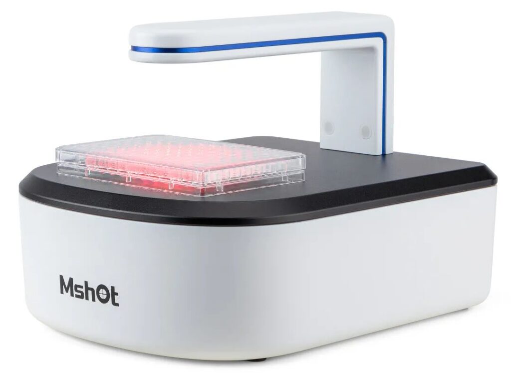
The compact Mshot Live Cell Imager operates stably within incubators for extended durations. Its client-server architecture enables remote control:
- Real-time experiment monitoring via mobile/PC avoids frequent clean bench access.
- Automated email alerts for abnormalities (e.g., sudden cell state changes or environmental deviations).
- Scheduled imaging and time-lapse video synthesis eliminate researcher attendance.
02 Red-Light Illumination: Low Phototoxicity for Long-Term Observation

Equipped with 625nm long-life, low-phototoxicity LED lighting, the system reduces photobleaching effects by >70% compared to conventional white light. This enables:
- Continuous wound healing tracking beyond 72 hours without compromising cell viability.
- Enhanced image clarity for adherent cells via high-penetration red light, ensuring precise edge detection even at high confluence.
03 End-to-End Data Acquisition for Wound Healing Assays
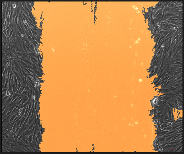
Post initial setup, the system automatically:
- Captures scratch regions at user-defined intervals with auto-focus and edge recognition.
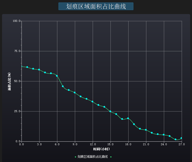
- Generates quantitative analyses (cell confluence, wound closure percentage) and graphical reports.
- Exports cell migration videos for presentations or publications.
We believe that the power of experimental data should not stop at “taking a few pictures”, but should extend to the insight into the laws behind cell behavior!