In the journey of exploring the microscopic world, scientists have long faced a core challenge: how to simultaneously observe multiple distinct structures within a sample. Traditional microscopy often resembles a black-and-white photograph—clear but limited in the scope of information it provides. The emergence of multichannel synthesis technology, however, is like giving the microscope “color vision,” allowing us to distinctly identify and localize multiple biological molecules in a single image.
What is Multichannel Synthesis?
Multichannel synthesis is a technique that merges image data from different “channels” into a composite image. In fluorescence microscopy, each “channel” corresponds to a specific fluorescent label. The core steps involved include selective labeling, channel-specific image acquisition, synthesis, and pseudocolor rendering.
Four Core Applications of Multichannel Synthesis
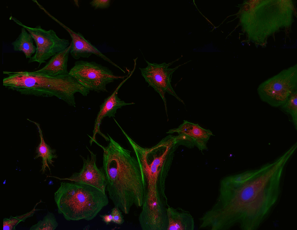
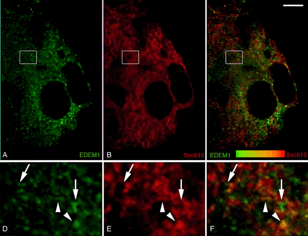
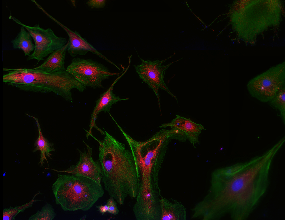
1. Precise Co-localization Analysis: Answering "Are They Together?"
By synthesizing images of two target proteins (e.g., protein A labeled in red, protein B labeled in green), the spatial relationship between them can be visually assessed. If the positions of both proteins coincide perfectly within the cell, the composite image will appear yellow, suggesting a potential interaction or co-localization in the same organelle. This provides direct visual evidence for studying protein interactions and signal transduction pathways.
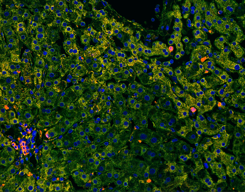
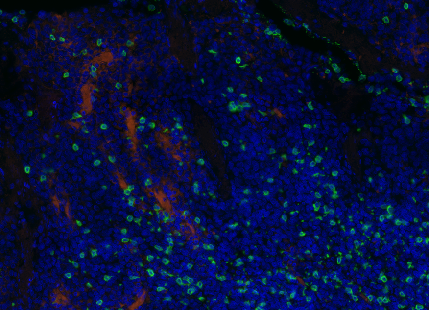
2. Efficient Multi-parameter Cellular Phenotyping: Answering "What Is It?"
In complex cellular populations (e.g., tumor microenvironments, immune cells), it is necessary to detect multiple biomarkers to accurately identify cell types. This is achieved by using specific surface or intracellular markers (e.g., CD3, CD4, CD8 for T cells) for multiplex labeling and synthesis. Researchers can differentiate distinct cell subgroups within a single image, enabling precise classification and counting of complex samples. This technique is widely used in immunology, stem cell research, and cancer studies.
3. Tracking Dynamic Processes: Answering "How Does It Change?"
When combined with live-cell imaging, multichannel synthesis allows tracking of dynamic processes of biomolecules.
Video: Fluorescent Changes Under MCS31
4. Pathological Diagnosis and Drug Screening: Serving Precision Medicine
Multichannel synthesis is a powerful tool for clinical diagnosis and drug development.
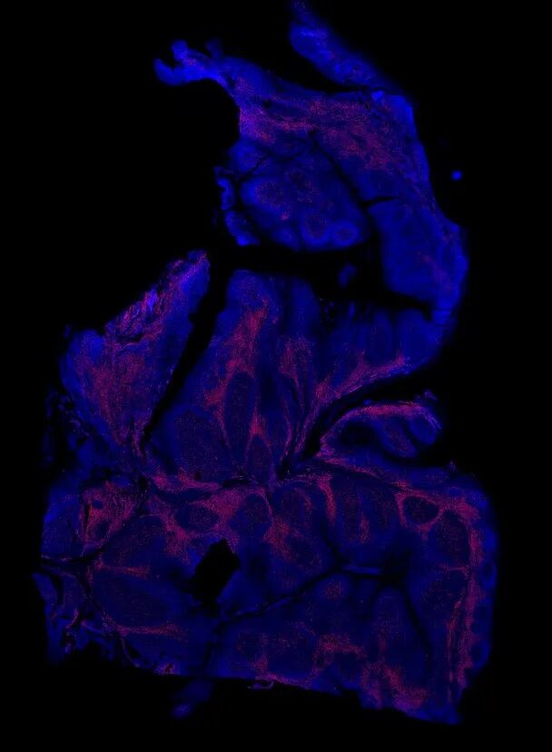
Clinical Diagnosis: In tumor pathology, multiplex immunofluorescence can simultaneously detect key prognostic markers such as HER2, ER, PR, and Ki-67 on a single tissue section, enabling precise molecular typing in one go, guiding targeted treatments.
Multiplex Immunofluorescence Observation
Challenges in Multichannel Synthesis Technology
Channel Crosstalk: Overlapping emission spectra of different fluorescent dyes can lead to signal “leakage,” causing false-positive co-localization results, which is a core issue affecting accuracy.
Image Registration: Color distortions from different wavelengths can prevent precise alignment of channel images. Calibration through hardware or software algorithms is necessary to correct these misalignments.
Photobleaching and Phototoxicity: Exposure to light can cause fluorescence signals to fade and damage living cells, posing significant challenges for long-term live-cell imaging.
Sample Autofluorescence: Background noise produced by the sample itself can lower the signal-to-noise ratio, interfering with the detection of weak signals.
MSHOT Solutions
1. Co-localization Analysis
Recommended: MSHOT’s Research-grade Fluorescence Microscope MF43-N, equipped with high-power LED light sources and a selection of filter combinations, supports up to six fluorescence channels. Its high image alignment precision ensures maximum excitation efficiency and minimal cross-talk between channels. When paired with MSHOT cameras (MSH12-BI/MSH20), high quantum efficiency and low readout noise ensure high-quality fluorescence signals even under low-light conditions, avoiding errors due to weak signals or noise.
(1) Recommended: MSHOT’s Research-grade Fluorescence Microscope MF43-N with a wide range of filter options supports up to six fluorescence channels. When paired with MSHOT’s MSX11/MSX3 cameras, it can effectively meet the need for multiplex labeling and imaging of immune markers such as CD3, CD4, and CD8.
(2) For large samples like tumor tissue sections, MSHOT’s Multiplex Fluorescence Digital Slide Scanning System MES200, paired with AI analysis software, provides high-definition panoramic scans of entire tissues and allows automatic identification, counting, and classification of cells. This facilitates precise quantification of cells.
3.Tracking Dynamic Processes & Drug Screening
Recommended: MSHOT’s Live Cell Imaging System MCS31
Non-invasive long-term observation in incubators, supporting time-lapse and multi-channel combinations. This allows multi-time point images to be synthesized into dynamic videos, visually presenting intracellular processes.
Fully automated, rapid high-throughput scanning of multi-well plates, enabling simultaneous testing of numerous drug sensitivity assays with multiple parameters, greatly accelerating the experimental process and improving both efficiency and accuracy.
625nm red-light technology significantly reduces phototoxicity and photobleaching, minimizing cell damage and ensuring physiological authenticity.
Conclusion
Multichannel synthesis technology is the key to unlocking the mysteries of the microscopic world. MSHOT, through providing stable, intelligent, and integrated imaging solutions, enables researchers and medical professionals to effortlessly harness this technology, transforming complex molecular data into intuitive, reliable color images. This ultimately aids in more precise scientific discoveries and clinical diagnoses.