
Mycobacterium tuberculosis
Applications of Mycobacterium tuberculosis
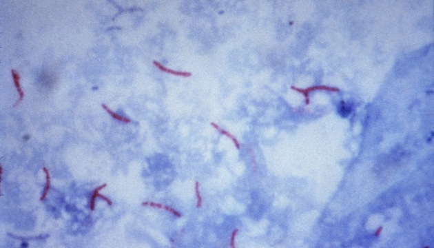
There are various methods for diagnosing tuberculosis, including smear microscopy, bacterial culture, genetic testing, and immunological testing. Smear microscopy is still widely used due to its simple operation but has a low detection rate. Bacterial culture offers high specificity but is time-consuming. Genetic testing features high accuracy and belongs to molecular-level detection. Immunological testing assists diagnosis through antibody detection. Among these, the fluorescent staining method is gradually gaining popularity in clinical applications due to its high detection rate and efficiency, outperforming traditional acid-fast staining with bright-field microscopy.
Pain Points in Mycobacterium Tuberculosis Detection
Bright-field microscopy suffers from low efficiency, while fluorescence microscopy is inconvenient to use and has high equipment requirements.
Bright-field acid-fast staining provides low detection rate and efficiency; fluorescent staining is recommended as the priority option.
Traditional fluorescence microscopes have inconvenient light sources; upgrading to LED or four-channel light sources is suggested to improve convenience and service life.
Fluorescence filter blocks must match the dyes. For Auramine O testing, the B2 Blue filter block is recommended, covering 425–475 nm excitation and emission above 495 nm to achieve better fluorescence performance.

Solutions for Mycobacterium Tuberculosis Detection
Bright-field Acid-fast Staining Solution: Choose the ML31/ML51-N microscope, featuring high-definition imaging, ergonomic design, and optional fluorescence upgrade.
● Fluorescence Microscopy: MF31 and MF23-M series, equipped with both bright-field and fluorescence functions, compatible with Auramine O, and high-sensitivity cameras for capturing weak fluorescence.
● Upright Microscope Upgrade Solution: Single/Triple fluorescence modules with long-life LED light sources, without affecting original functions.
● Fluorescence Microscope Upgrade Solution: Four-channel MG-120 or wide-spectrum MG-100, LED light sources with adjustable brightness, wide compatibility, and convenient touchscreen operation.