

In fields like cell biology, immunofluorescence, and tumor research, high-quality fluorescence imaging is often the key to revealing biological phenomena. However, many researchers face imaging challenges in their pursuit of perfect images—either the fluorescence signal is too weak to visualize, or excessive noise obscures key signals.
These issues are usually not due to sample preparation or the equipment itself, but rather stem from several critical imaging setup factors.
Today, we’ll share five essential points for fluorescence imaging. Mastering these will ensure you never have to worry about failing to capture journal-quality images again!
1. Sample/Slide Thickness
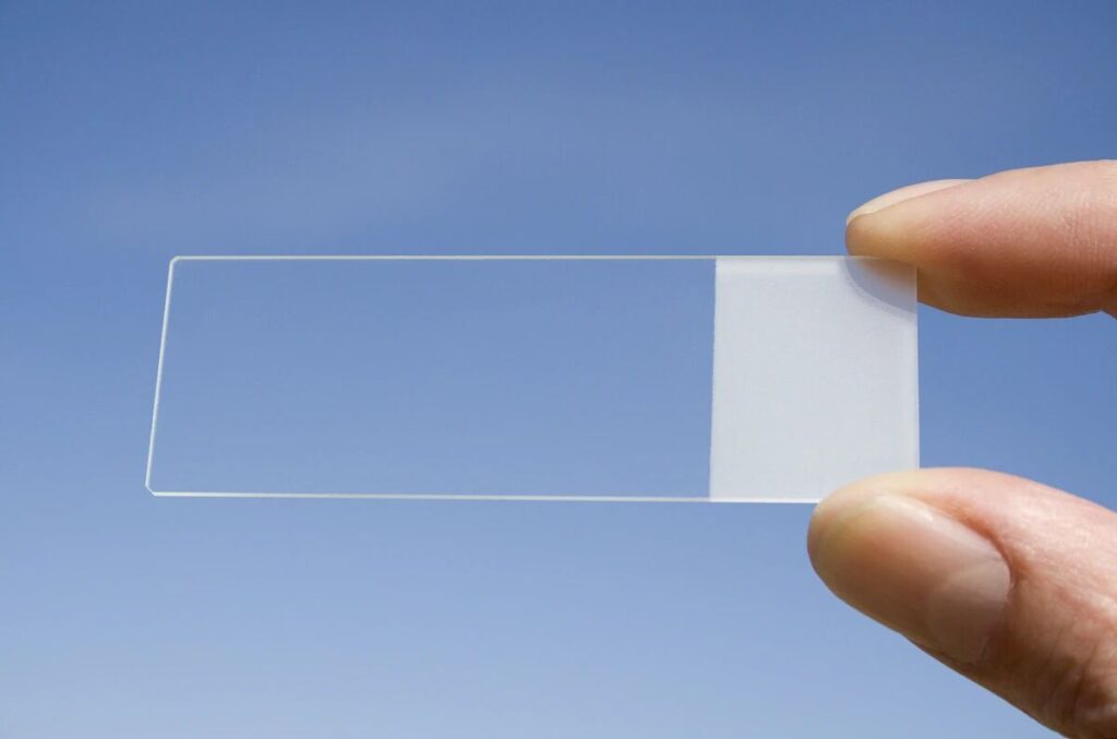
Including slides, cover glasses, and tissue sections should not be too thick. Thick slides absorb more light and fail to allow excitation light to properly focus on the sample. Thick tissue sections can cause cell overlap or the presence of impurities, which can obscure key observations.
Recommendations/Tips:
- The thickness of the slide should be between 0.8 to 1.2 mm, and the cover glass should be approximately 0.17 mm.
- For enhancing excitation light, you can use interference cover glasses. These specialized cover glasses are coated with multiple layers of materials (e.g., magnesium fluoride) that interfere with different wavelengths of light, enabling fluorescence to pass through smoothly.
2. Proper Filter Set Matching: The Key Factor
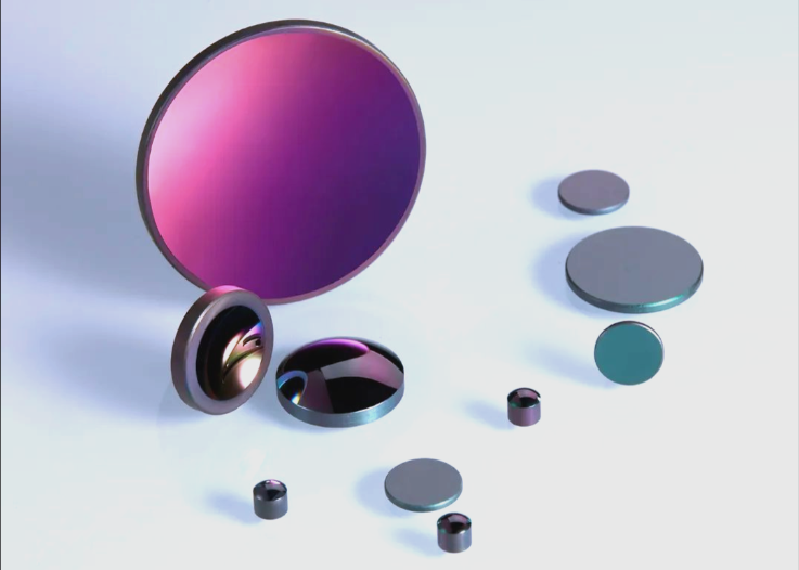
Different fluorescent dyes have distinct excitation/emission spectra. Using the wrong filter set is essentially pointless.
Recommendations/Tips:
- Verify the dye’s spectral range and select the corresponding filter set (e.g., FITC: EX490/EM520 nm).
- Particularly for multi-channel co-staining samples, avoid spectral overlap to minimize crosstalk.
3. Fluorescence Quenching Protection

Fluorescent signals often fade too quickly during imaging, typically due to fluorescence quenching caused by exposure to light. This not only affects image quality but may also interrupt experiments and result in data loss.
Recommendations/Tips:
- Use mounting media that contains DABCO or similar anti-quenching agents.
- Try to keep the entire process in the dark, especially during staining, mounting, and transfer stages.
- Capture channels prone to quenching (e.g., Cy5) first, followed by more stable channels like DAPI.
4. Have You Chosen a Fluorescence Objective Lens?
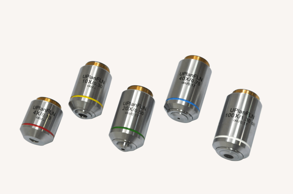
A fluorescence objective lens, as the name suggests, is designed specifically for fluorescence observation. These lenses use fluorite (Fluorite) glass, offering better optical performance compared to standard lenses, resulting in brighter, higher-contrast images.
Recommendations/Tips:
- Pay attention to the Numerical Aperture (NA) of the objective lens: the higher the NA, the better the lens’s light-gathering ability and resolution. For fluorescence imaging, choose an objective with a high NA.
5. Have You Chosen the Right Fluorescence Light Source?
The brightness and signal-to-noise ratio of fluorescence images are largely dependent on the strength and stability of the excitation light source. Incorrect light source selection can lead to weak signals, long exposure times, and enhanced quenching.
Recommendations/Tips:
- Prioritize high-power LED light sources: compared to traditional mercury or metal halide lamps, LED light sources have distinct advantages.
- Brightness & Stability: LED sources instantly reach full power, offering stable output without the gradual dimming seen with traditional bulbs, ensuring reproducibility in experimental results.
- Long Lifespan & Low Maintenance: LED lights last tens of thousands of hours, far exceeding traditional bulbs, reducing long-term usage and maintenance costs.
- Quick Switching & Flexible Control: LEDs allow for millisecond-level on/off switching, ideal for multi-field and time-point live cell imaging, minimizing unnecessary sample exposure.
Product Recommendations
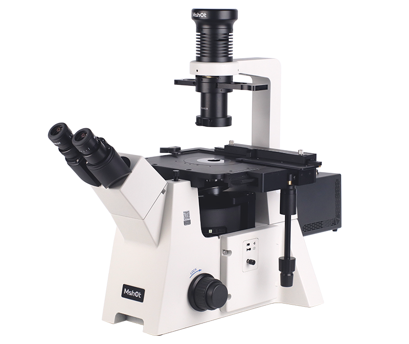
MSHOT MF53-N Inverted Fluorescence Microscope is a classic research-grade inverted fluorescence microscope, featuring an exceptional infinity-corrected optical system and a long-working-distance semi-apochromatic objective lens. It is equipped with MSHOT high-power LED light source, the MG-100, which covers a broad spectrum.
The microscope can be configured with a variety of filters (UV, V, B, G, Y, R) to observe common fluorescent dyes like DAPI, FITC, TRITC, and Alexa Fluor series, widely used in CTC detection, FRET, and cell immunology. It is commonly applied in university research labs, research institutes, and clinical departments such as pathology and laboratory medicine.
Its 6-position rotating fluorescence accessory design allows for quick and precise filter changes, minimizing crosstalk and enhancing multi-color imaging efficiency. The stable optical structure ensures high consistency in image acquisition.
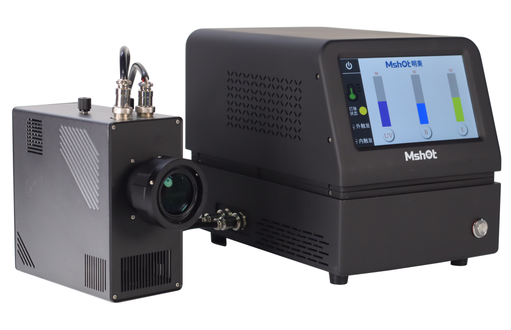
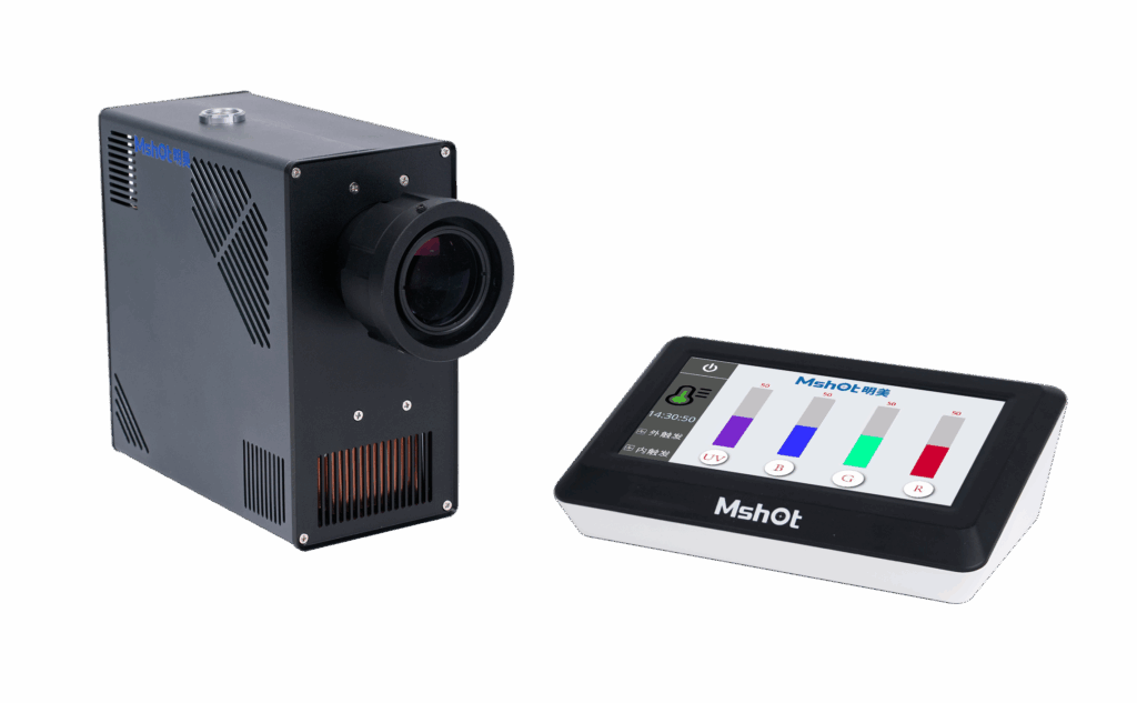
The MG-150 is a high-efficiency, long-lifetime fluorescence light source that achieves more than double the light efficiency of traditional mercury lamps in the RGY excitation bands.
- Instant on/off capability with 3 independent channels for precise brightness adjustment (0-100% for UV, B, Y channels).
- Smart brightness memory function for each channel to minimize fluorescence quenching.
- High compatibility with research-grade fluorescence microscopes from leading brands.
Real Research Case
In a recent study on hypertension published in the Journal of Extracellular Vesicles, a team from China Pharmaceutical University used MSHOT MF53-N Inverted Fluorescence Microscope to conduct cell migration imaging, successfully revealing new mechanisms of vascular remodeling and identifying potential therapeutic targets!
High-quality fluorescence imaging results from a combination of precise sample preparation, accurate filter matching, comprehensive quenching protection, high-performance objective lenses, and stable excitation light sources. Systems like the MSHOT MF53-N and MG150 integrate these key elements to provide stable, reliable support for high-level research in your laboratory.
We believe excellent scientific discoveries start with reliable data. If you want to learn more about optimizing your fluorescence imaging platform, feel free to contact us for customized solutions and detailed technical information tailored to your experimental needs.