Beneath the lens of a microscope, the magical microscopic world unfolds before us, where countless vivid “little stories” lie hidden. Today, let’s embark on a journey together to explore these fascinating “biological wonders”!
(Note: The following images are sourced from the Internet)
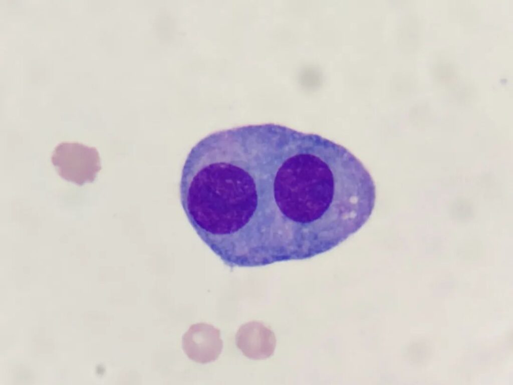
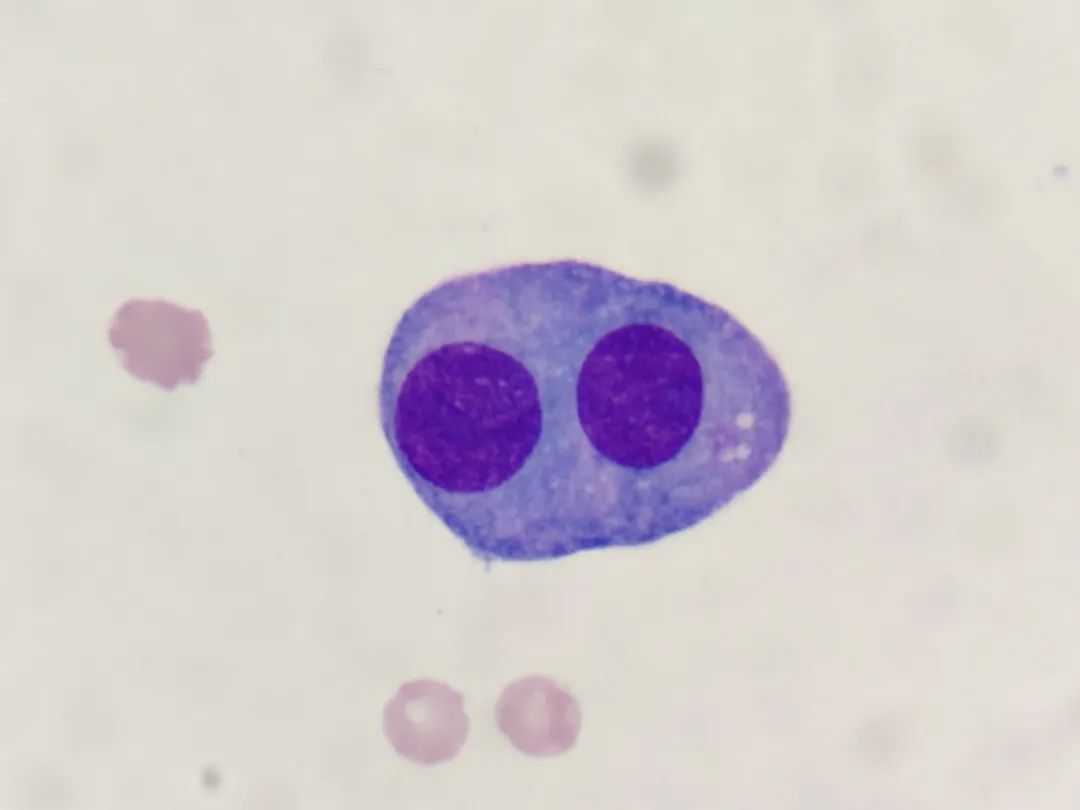
Mesothelial cells line the surfaces of body cavities such as the pleura, peritoneum, and pericardium, primarily functioning to protect and lubricate. These cells are flat and membranous, reducing friction between organs. Under the microscope, the two nuclei of this mesothelial cell resemble a “double-yolk egg,” creating a unique visual effect that is both scientific and somewhat warm.
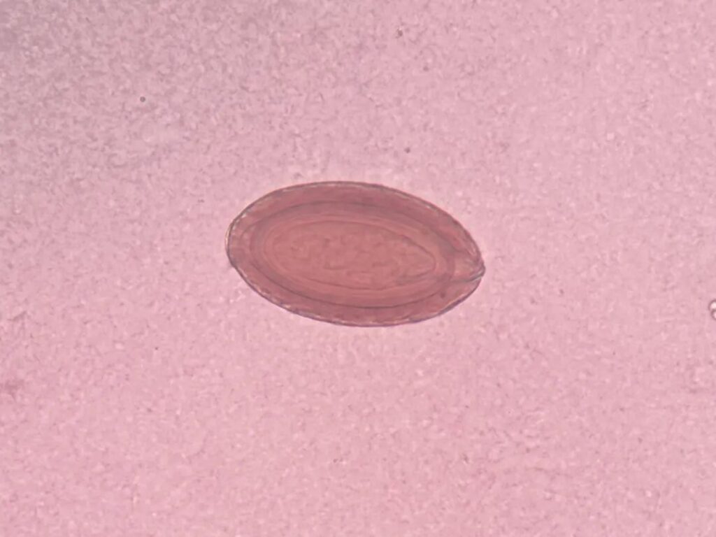
2.Acanthocephalus dirus Eggs: “Almonds” in the Microscopic World
Acanthocephalus dirus is a type of parasite, commonly found in the small intestines of pigs, where it affects the animals’ health. The eggs of this parasite appear oval under the microscope, with their hard shells looking like tiny “almonds.” Though they are the products of a parasite, they have a unique charm, showcasing the rich diversity of the biological world.
Calcium oxalate crystals are usually present in human urine, formed by metabolism in the body after consuming foods rich in oxalic acid. Under the microscope, the yellowish-orange calcium oxalate crystals resemble mini dumbbells, being crystal clear, interesting and lovely, like “little artworks” in the microscopic world.
4.Platelet Aggregation: A Spectacular “Coagulation Landscape” Like a Galaxy
Platelets are tiny “coagulation guardians” in the blood, responsible for hemostasis and coagulation. At the site of a damaged blood vessel, platelets quickly aggregate and adhere to the vessel wall, forming a platelet plug that looks as brilliant as a galaxy. Such vitality and order make one can’t help but marvel at the microscopic beauty of life.
5.Fat Droplets in Prostatic Fluid: Brightly Refractive “Little Planets”
Under the microscope, the fat droplets in prostatic fluid have extremely strong refractivity. After staining, they present vivid colors, resembling floating little planets. This scene enriches our understanding of the body’s microscopic structures and also makes people feel a little more in awe of the complexity of life.
6.White Blood Cells vs. Bacteria: A “Life-and-Death Struggle” Without Gunsmoke
As the main force of the immune system, white blood cells are always on guard against foreign invaders. Under the microscope, white blood cells are engaged in a fierce “life-and-death battle” with bacteria. The two sides are evenly matched, and the outcome remains undecided. This “microscopic battle” makes people feel the vitality of life and the tenacity of white blood cells…
The world under the microscope is colorful, and every image tells a story of life. There are many more such stories unfolding all the time. For those who are interested, you can get hands-on yourself—using a biological microscope allows you to admire these wonderful microscopic worlds.
We recommend Mshot biological microscope ML51-N:
It adopts excellent infinite-distance independent achromatic optics, paired with a wide-field eyepiece, delivering clear imaging and vivid colors. It is an excellent helper for morphological observation and detection.