
In the microscopic universe of the human brain, microglial cells act as an elite “immune defense” force, tirelessly protecting our nervous system 24/7. These primitive macrophages, originating from the embryonic yolk sac, are not only responsible for clearing dead cells and pathogens but also play a critical role in key processes of brain development—regulating neurovascular genesis, pruning synaptic connections, and even influencing the fine-tuning of neural circuits.
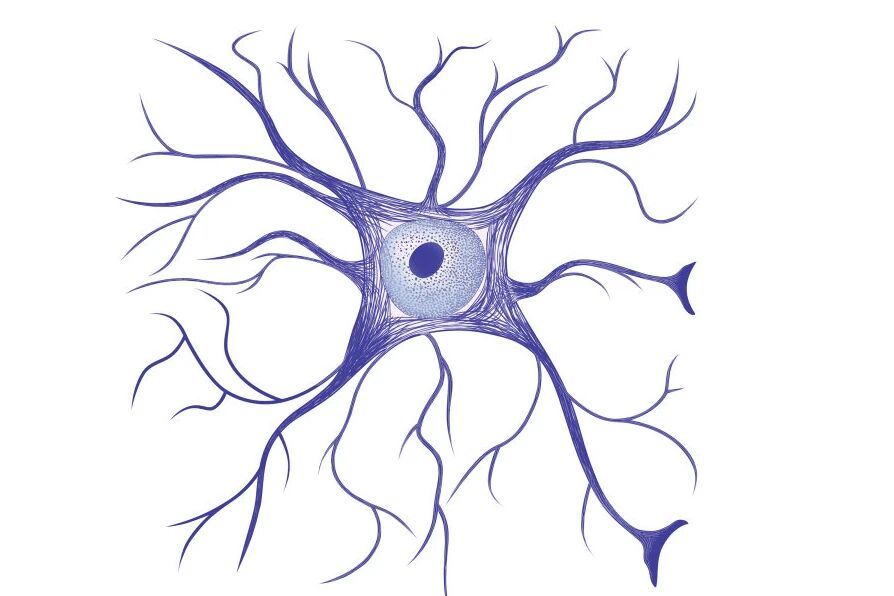
Image 1: Detailed branching structure of microglial cells in brain tissue
The morphology of microglial cells is highly plastic and closely related to their functional state. In normal brain tissue, microglial cells exhibit a highly branched structure with three to four levels of branching, and there is minimal overlap between branches of adjacent cells. These branched microglial cells are often referred to as “resting microglial cells.”
Surprisingly, the “resting state” of microglial cells, as traditionally understood, is actually a highly active monitoring state. Recent research has shown that these cells make direct contact with neuronal synapses every hour to monitor synaptic function. This dynamic surveillance system activates the microglial cells to transform into phagocytic cells upon detecting inflammation, infection, or injury, thus initiating immune responses.
However, scientific research has long been hindered by technical limitations:
- Traditional two-dimensional cell cultures cannot replicate the brain’s three-dimensional microenvironment;
- Animal models struggle to reproduce the human-specific neurodevelopmental processes;
- Static observation techniques fail to capture key dynamic biological processes.
1. Brain Organoids: Opening a New Era of 3D Research
In 2013, the Lancaster team made a groundbreaking achievement by cultivating the first multi-brain region organoid using a rotating bioreactor, successfully reproducing neurogenesis and neuronal migration in vitro.
In 2022, the Pasca team took this further by successfully transplanting human brain organoids into the brains of newborn rats, establishing a functional integrated human-mouse hybrid neural network. This marked the shift of organoid technology from static simulation to dynamic functional research.

Image 2: Potential applications of human stem cells
The emergence of organoid technology has provided a new path for addressing the challenges in microglial cell research. These “miniature brains,” self-assembled from induced pluripotent stem cells (iPSCs), can closely simulate the complex processes of human brain development, offering an ideal platform for studying interactions between microglial cells, neurons, astrocytes, and other cell types.
2. The Overcoming Technological Challenges
Despite progress in brain organoid technology, researchers still face three major challenges:
①Culturing and Monitoring Difficulties: Organoids require precise control over the composition of the culture medium, gas concentration, and temperature, while the strong light emitted by traditional microscopes can interfere with normal cell growth.
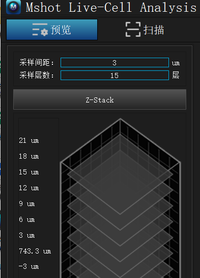
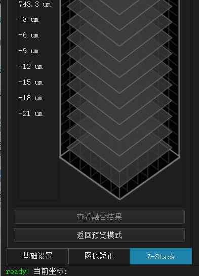
②3D Imaging Challenges: The complex three-dimensional structure of organoids makes it difficult for conventional microscopes to achieve clear, all-around imaging.
③Dynamic Quantification Challenges: The phagocytosis, migration, and secretion processes of microglial cells are dynamic and variable. Traditional methods cannot achieve long-term continuous observation or quantitative analysis.
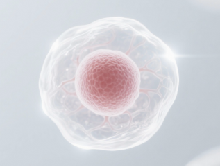
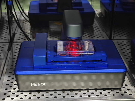
Image 3: Cells bleached under high-intensity light
Precision Medicine
Patient-derived organoids (PDOs) retain the genomic and epigenetic features of original tumors, making them an excellent platform for personalized drug testing. For ovarian cancer treatment, doctors can culture patient-specific organoids within 2-4 weeks to test responses to different chemotherapy regimens. Success rates have increased from 23-53% with traditional methods to 85% using deep learning predictions.
Rare Disease Research
Over 7,000 rare diseases lack treatment options, with only about 400 being studied. A major bottleneck is the lack of suitable animal models. Organoid technology, by directly using patient cells to construct disease models, brings hope for these “orphan diseases.”
Thus, compared to traditional research methods, organoids offer higher efficiency and more accurate data.
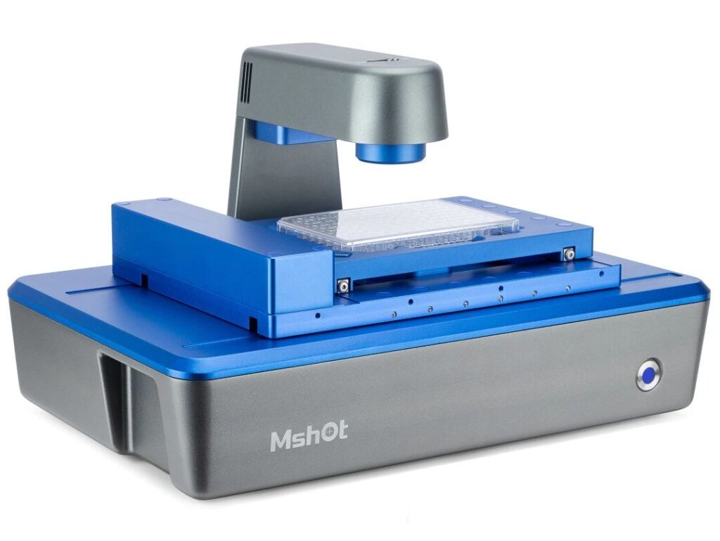
3. MSHOT MCS31: A Breakthrough Technological Solution
To address these challenges, MSHOT has introduced the MCS31 live cell imaging system, offering a comprehensive solution:
Ultra-low Light Toxicity Imaging Technology
The MCS31 uses a 625nm red LED light source, reducing photon energy and lowering light toxicity to less than 30% of traditional equipment. This breakthrough technology extends continuous observation time from hours to days, allowing researchers to observe the entire activation process of microglial cells without interference.
AI Smart Analysis Platform
The built-in deep learning algorithm automatically identifies and tracks the dynamic behavior of microglial cells, quantifying the number and size of phagocytic vesicles in real-time, analyzing cell migration trajectories and speeds, and generating growth curves and statistical reports.
Multi-Scenario Adaptability
The MCS31 supports a variety of container types, including 6-well to 384-well plates, culture bottles, and dishes, making it compatible with different organoid cultivation and observation needs.
4. Breakthrough Applications of MCS31 in Scientific Research
The MCS31 has demonstrated immense value in multiple research areas:
Neurodevelopment Research
It enables continuous observation of microglial cell involvement in synaptic pruning for up to 72 hours, capturing dynamic images of “synaptic phagocytosis” for the first time, providing direct evidence for understanding the fine-tuning mechanisms of brain neural networks.
Disease Mechanism Research
In Alzheimer’s disease models, researchers observed microglial cells clearing β-amyloid plaques and discovered functional heterogeneity in microglial cells with different activation states.
Drug Screening Platform
MCS31 enables high-throughput organoid drug screening, automatically analyzing the effects of drugs on microglial cell functions, greatly improving the efficiency of neuropharmaceutical research.
5. The Revolutionary Significance of MCS31 Technology
The Mshot MCS31 brings not only improvements in technical parameters but also a shift in research paradigms:
- From “end-point observation” to “process monitoring”
- From “2D static” to “3D dynamic”
- From “qualitative description” to “quantitative analysis”
- From “manual operation” to “intelligent automation”
This breakthrough allows researchers to achieve “visualized” research of life processes, rather than merely capturing snapshots of life.
Mshot MCS31 is not just an instrument; it is the key to unlocking the mysteries of the brain. It enables researchers to “see” processes that were once invisible, “quantify” changes that were previously immeasurable, and “understand” phenomena that were once unexplained.
In the quest to uncover the mysteries of the brain, we need not only clearer images but also a deeper understanding of dynamic life processes.
Mshot MCS31 is turning this goal into a reality, driving brain science research into a new era.