As the global infectious disease prevention and control situation becomes increasingly complex, the importance of the CDC virus laboratory in the field of public health becomes increasingly prominent, playing an irreplaceable and key role in infectious disease prevention and control. Optical microscopes are widely and deeply used in the CDC virus laboratory. They play an irreplaceable role in virus detection, mechanism research and prevention and control support, providing strong support for the development of virology and public health prevention and control.
1.Application of microscope in virus laboratory
The CDC Virus Laboratory is a core technical support platform for infectious disease prevention and control, pathogen research, and public health emergency response. Microscopes, as basic laboratory tools, play an irreplaceable role in virus detection, mechanism research, and prevention and control decision-making.
|Virus Detection and Diagnosis
The respiratory seven-in-one test/nine-in-one test uses immunofluorescence technology to detect fluorescent signals through a fluorescence microscope. It can quickly and accurately detect a variety of respiratory pathogens, provide doctors with timely etiological information, help formulate reasonable treatment plans, improve patients’ cure rate and recovery speed, and reduce the risk of disease transmission.
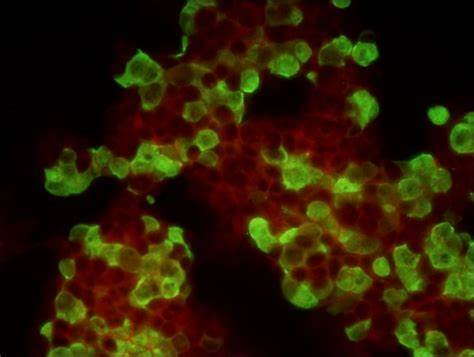
Immunofluorescence of seven respiratory tests taken by MSHOT MF31 (green represents viral reaction)
| Observation of virus morphology and structure
Virus invasion causes pathological changes in cells or tissues, and the pathological morphology of cells or tissues stained by IHC or HE can be observed under bright field microscopy.
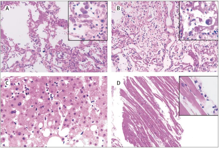
Pathological tissue sections of patients with severe pneumonia infected with SARS-CoV-2 (picture from the Internet)
|Cell Culture Epidemiology (CPE)
Initial screening with an inverted microscope: observe cell morphology changes daily and record the progression of lesions (such as vacuolization and shedding) Fluorescence microscopy analysis: use fluorescently labeled antibodies or dyes to accurately locate the virus-infected area
Cells infected with rotavirus (green) have begun to vacuolate
|Virus-host interaction research
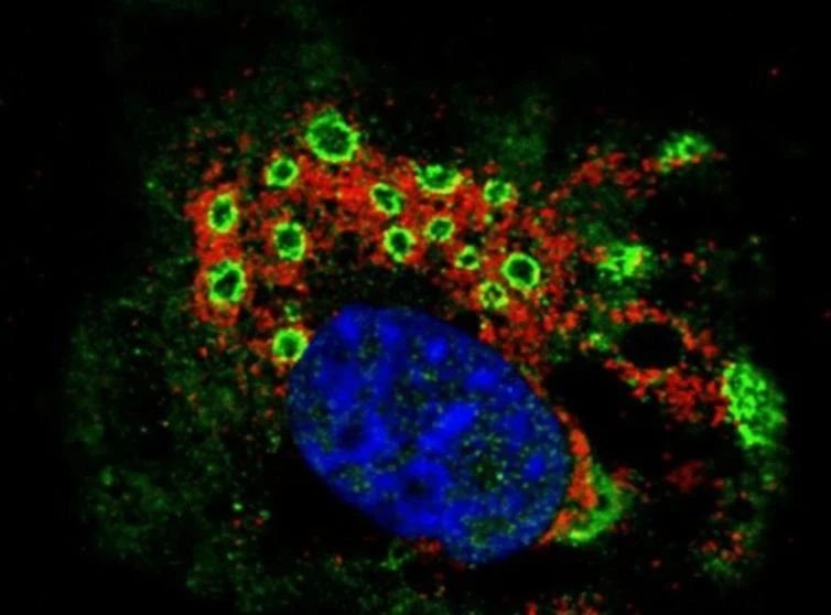
Fluorescence in situ hybridization (FISH) technology: By hybridizing fluorescently labeled nucleic acid probes with the viral genome and detecting fluorescent signals under a fluorescence microscope, the viral nucleic acid can be accurately located and detected, providing an important basis for early diagnosis of viral infection and disease monitoring.
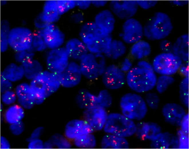
FISH images taken by MSHOT MF43-N
2.Challenges of microscopic imaging in virus observation
The size of a virus is usually less than 100nm, far exceeding the resolution limit of an optical microscope (about 200nm), so it is difficult to directly and clearly observe the morphology and structure of the virus with an optical microscope. In practical applications, conventional detection relies on indirect methods: observing the pathological morphological changes in host cells caused by viral infection (such as vacuolization, syncytium formation) or tissue damage characteristics to preliminarily judge the infection status.
If fluorescence microscopy is used for detection, viral components can be located at the subcellular level through specific fluorescent labeling strategies (such as immunofluorescent antibody labeling of viral antigens, or nucleic acid probe labeling of viral genomes). However, due to the small size and low copy number of viral particles, the intensity of the fluorescent signal generated by the labeling is significantly limited. To overcome this problem, high-intensity excitation light and a highly sensitive microscope camera are required for imaging to enhance the capture and recording of signals, thereby improving the accuracy and sensitivity of virus detection.
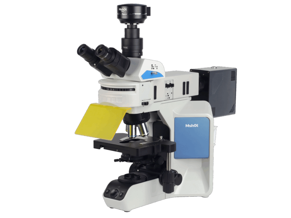
3.MSHOT Microscope Solutions
1) Respiratory tract seven-in-one test/nine-in-one test plan
Recommended upright fluorescence microscope MF31/MF23 + microscope camera MD60/MDX10 (main recommendation): suitable for fluorescence observation of respiratory tract seven-joint test and nine-joint test, B excitation fluorescence (460-490nm) shows green fluorescence reaction
2) Routine observation protocol for virus-infected tissue sections and immunohistochemical sections
Recommended upright biological microscope ML51-N/ML45/ML31+microscope camera MDX10/MD60, etc.
3) Cell culture lesion (CPE) observation plan
Recommended: ① Inverted fluorescence microscope MIX60-FL (three colors) + MSX3/MDX10; ② Inverted fluorescence microscope MF53-N + MSX11-C
4) Fluorescence in situ hybridization (FISH) technology detection plan
Recommended: MF43-N + wide spectrum high power light source MG-100/MG-120 + high sensitivity camera MC50-S/MS23/MSX3 + FISH software