Why is the background not dark during fluorescence observation?
In the lab, the sample under a fluorescent microscope should appear on screen like a starry sky—bright target signals against a pitch-black background.
But in reality, many researchers face the headache of “grayish backgrounds” and “stray light interference”:
fluorescence signals seem veiled
contrast drops
“false positives” pop up
What’s causing this?
Blame it on sample prep flaws, equipment calibration issues, or invisible environmental nuisances.
Today, we’ll break down the root causes of fluorescent background noise from a pro perspective—and share practical fixes to restore your pristine fluorescence view.
Cause Analysis: Four Dimensions to Pinpoint Background Noise Sources
1. Sample-Related Issues
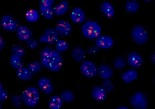
Fluorescent background “noise” often stems from the sample itself. Autofluorescence is the primary culprit: certain fixatives (e.g., paraformaldehyde), mounting media (glycerol), or glass slides (containing silicates) can emit non-specific fluorescent signals when illuminated by excitation light. Excessive fluorescent probe concentration may cause self-quenching or bind to non-target molecules, creating diffused background—for example, DAPI over-staining often results in blue cytoplasmic interference.
2. Environmental Interference
External environments serve as “stealth channels” for stray light. Ambient light leakage (e.g., room lighting or poor equipment sealing) directly contaminates the imaging light path, especially noticeable during long exposures with high-sensitivity cameras. Additionally, temperature fluctuations can cause thermal expansion/contraction of optical components, altering light path collimation and inducing stray reflections.
3. Equipment Performance Limitations
The hardware configuration of fluorescence microscopes directly affects signal-to-noise ratio. Inadequate filter set matching is a common issue: if excitation and emission filters have overlapping bands (e.g., too wide bandwidth or insufficient cut-off depth), excitation light can “leak” directly to the detector. Moreover, objectives with excessively high numerical aperture (NA) may enhance resolution but also collect more off-axis stray light (e.g., Rayleigh scattering).
4. Operational Parameter Pitfalls
Even with intact equipment, improper parameter settings introduce background noise. Excessive excitation light intensity aggravates photobleaching and autofluorescence; prolonged exposure amplifies camera dark current and read noise.
Solutions: Efficient Noise Reduction for Pure Dark-Field Backgrounds
Sample Optimization:Switch to low-autofluorescence reagents, control probe concentration, and store stained samples in the dark. Optimizing sample processing (e.g., using quartz slides with low autofluorescence and adjusting dye concentration) significantly reduces such interference.
Environmental Control:Operate in a darkroom and regularly calibrate the microscope’s light path to block external interference. Clean objectives and filters periodically to avoid dust scattering.
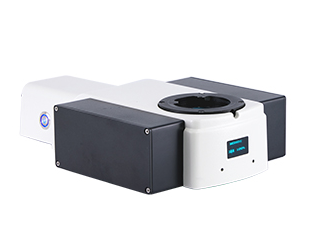
Equipment Upgrades:Select fluorescence modules and light sources with appropriate wavelength bands. Upgrade to high-resolution microscopy systems (e.g., MSHOT Four-Channel Light Source MG120 and upright digital fluorescence module) to suppress stray light targetedly.
MSHOT Four-Channel Light Source MG120
MSHOT Upright Digital Display Fluorescence Module
Parameter Adjustment:Debug parameters via software: use black balance, reduce dark areas, and balance brightness/exposure for optimal imaging effects.
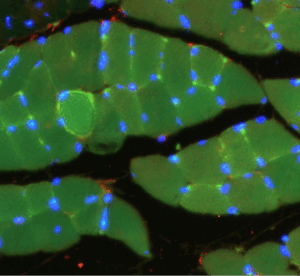
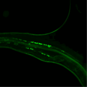
After hardware optimization and software debugging, the final fluorescent sample images feature:
Clear fluorescent markers with highvisibility
Dark background with high contrast
Small intestine tissue sections captured by Mshot MF43-N + MSX11
Root stem fungal invasion captured by Mshot MF43-N + MSX11
Precision Filtering: Equipped with imported narrow-band filter sets, excitation/emission light isolation >99.9% to eliminate spectral crosstalk.
Intelligent Noise Reduction: Paired with Mshot intelligent image recognition software algorithm for one-click weak signal optimization.
Stable Light Path: Fully sealed anti-vibration design, compatible with oil/water immersion objectives and high NA objectives, stray light coefficient <5%.
The “purity” of fluorescent imaging is a blend of science and art—from sample preparation to equipment debugging, every detail impacts result reliability. Choose professional equipment, optimize experimental processes, and let every fluorescent signal speak with precision.


")
