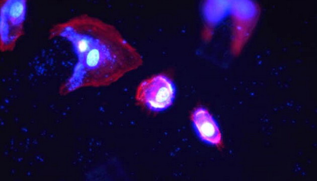
Live cell imaging is a powerful technique for investigating dynamic cell growth processes and the effects of various influencing factors, and it has been widely adopted across many different fields.Cells undergo dynamic changes in vivo. Observing and studying their growth processes and the effects of external controlled variables necessitates live cell imaging technology, which enables real-time recording of cell growth status, expression activities, and more.Additionally, cells can be fluorescently labeled according to research requirements to study dynamic events including embryonic development, cytoskeleton changes, and cell division.
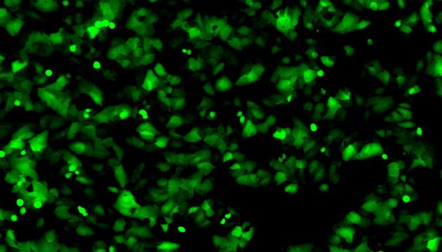
The primary significance of live cell imaging lies in monitoring cell culture conditions to evaluate culture status, experimental performance, and the optimal timing for cell passage.For example, it is used to calculate confluence in cell culture and assess the appropriate culture timing, as well as to measure cell migration in scratch assays.In addition, fluorescence transfection monitoring requires fluorescence microscopy to visualize uptake and expression.
Pain Points in Live Cell Imaging
Live cell imaging requires a balance between imaging quality and cell health. Attention must be paid to culture conditions, light source selection, the toxicity and compatibility of fluorescent dyes, and camera sensitivity.In addition, auto-focusing, constant temperature, and stable liquid volume control are required to reduce focal plane drift and ensure quality during long-term observation.
Solutions for Live Cell Imaging
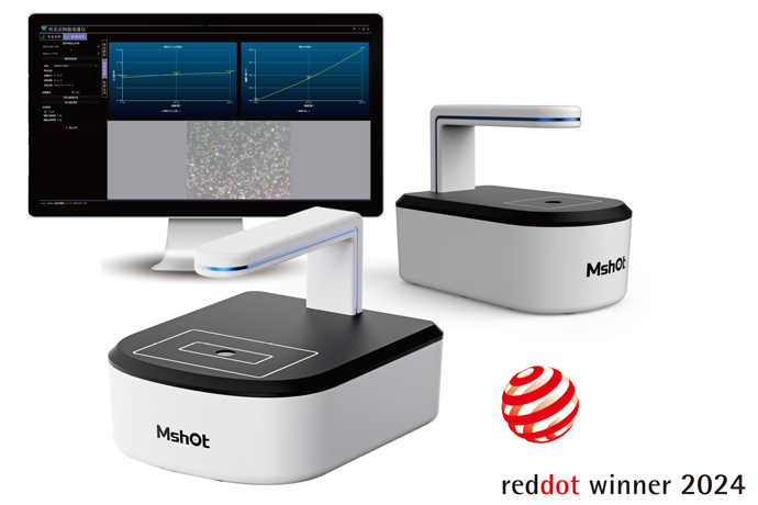
Primary Solution: MCS11/MCS21 Live Cell Imager. It can be placed inside an incubator for long-term observation, supports remote management, and automatically reports cell confluence. Compatible with brightfield and fluorescence imaging, it greatly improves the efficiency of live cell culture imaging and long-term experiments.
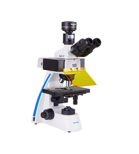
Traditional Solution: MI52-N or MF52-N fluorescence microscope with a high-sensitivity camera. Features multi-mode observation and low phototoxicity.
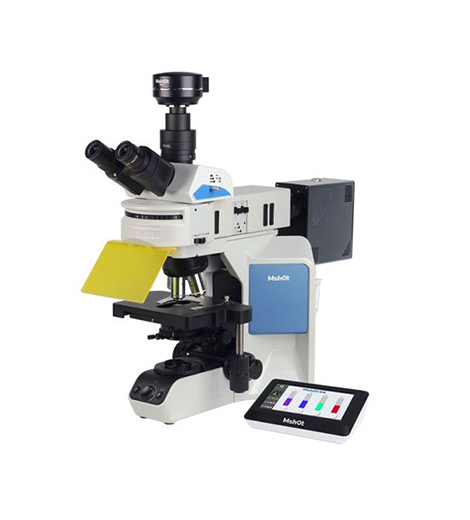
Research-Grade Solution: MCS31 Live Cell Imaging Scanner and motorized MF53-N research-grade microscope with a high-sensitivity camera. Equipped with a precise motorized stage, it supports brightfield and fluorescence imaging, enables multi-well scanning, and meets advanced research requirements.
LED Fluorescence Upgrade Kit: Compatible with most microscope brands. Combines an LED light source and high-sensitivity camera for cost-effective fluorescence observation.