
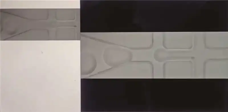
An inverted microscope is often a practical choice for transparent microfluidic chips with tubing and reservoirs above the sample. Fluorescence imaging enables the observation of labeled cells and fluorescence-based assays, while automated live cell imaging supports experiments that run overnight or across several days. For silicon molds and metal electrodes, reflected-light microscopy serves a different inspection role.
The right system must also accommodate the chip’s bottom thickness, sample holder, fluid connections, and required imaging speed.
This guide explains how to select a microscope for microfluidic experiments, using four MSHOT systems as examples: MJ31, MIX60, MIX60-FL, and MCS31.
Quick Comparison: Which Microscope Fits Your Microfluidic Experiment?
| Experimental task | Imaging requirement | MSHOT model to consider |
|---|---|---|
| Inspect silicon molds, metal electrodes, and chip surface features | Reflected-light imaging | MJ31 |
| Observe droplets, particles, flow behavior, and unstained cells | Brightfield and phase contrast | MIX60 |
| Image fluorescently labeled cells, particles, and assays | Fluorescence with brightfield or phase contrast | MIX60-FL |
| Monitor cell growth and responses over hours or days | Automated time-lapse and multichannel imaging | MCS31 |
Start with the experimental task, then check optical and mechanical compatibility. A microscope with the right imaging modes still needs to focus through the chip bottom and accommodate the complete experimental setup.
1. Microfluidic Chip and Mold Inspection: MJ31
For laboratories that fabricate their own microfluidic devices, microscopy begins before the first fluid experiment.
Researchers may need to inspect channel patterns on a mold, check inlet and outlet positioning, or examine patterned electrodes. These tasks have different requirements from imaging cells inside a transparent chip.
When reflected-light microscopy is useful
Silicon molds and metal structures may be opaque, making transmitted-light observation unsuitable. Reflected-light microscopy illuminates the sample from above through the objective, allowing accessible surface features to be examined.
The MSHOT MJ31 upright metallurgical microscope provides an infinity optical system and long-working-distance plan achromatic objectives for this type of inspection.
Its 204 × 204 mm stage travel range supports the examination of relatively large substrates and samples. Reflected-light illumination and polarization observation provide contrast options for suitable materials and surface features.
Typical inspection tasks
MJ31 can support the examination of:
Silicon mold surface patterns
Metal electrode structures
Visible surface contamination or fabrication defects
Inlet and outlet positioning
Accessible chip interface features
Bonding defects and enclosed channels require particular care. Their visibility depends on the transparency of the materials, the location of the feature, and the illumination method. A reflected-light image alone may not reveal every internal defect.
For laboratories producing their own chips, MJ31 can serve as a complementary fabrication inspection station before fluid testing begins.
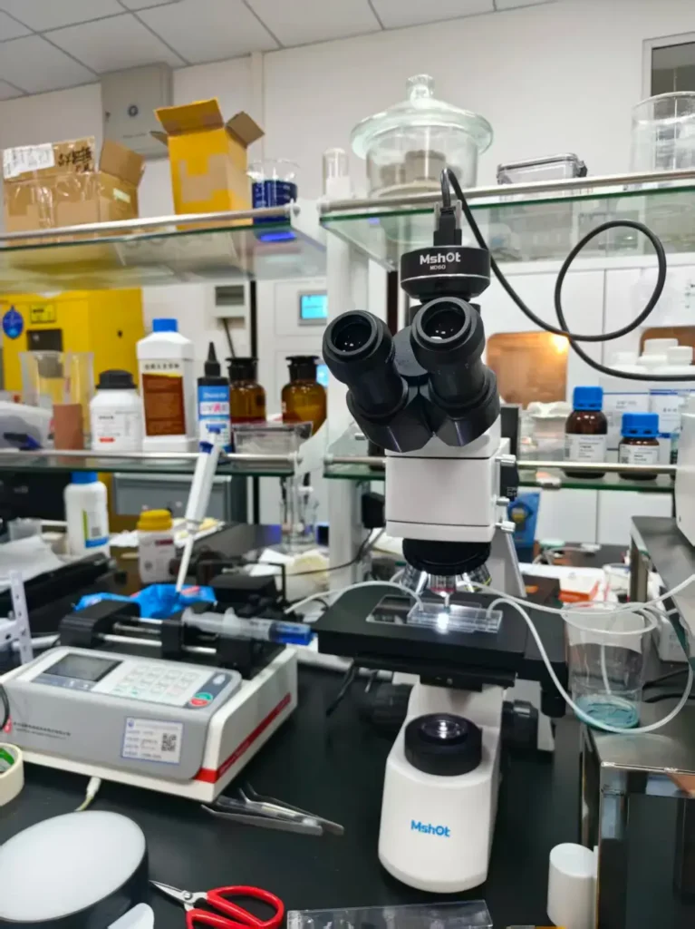
MSHOT MJ31 upright metallurgical microscope for reflected-light surface inspection.
2. Brightfield and Phase Contrast Imaging for Microfluidics: MIX60
For routine observation inside transparent microfluidic chips, an inverted microscope is often a convenient starting point.
The objective observes the sample from below, leaving the upper surface available for reservoirs, tubing, and experimental adjustments. This arrangement is useful when researchers need to change flow conditions while watching the sample.
Observing transparent samples
The MSHOT MIX60 inverted microscope supports brightfield and phase contrast imaging.
Brightfield can be used to observe visible channel boundaries, particles, and other features with sufficient contrast. Phase contrast improves the visibility of suitable unstained transparent specimens, particularly living cells.
This helps researchers inspect cell morphology and attachment without introducing fluorescent labels for every observation.
Accommodating chips and fixtures
MIX60 features long-working-distance phase contrast objectives, a flip-up stage design, and a universal sample holder.
The stage can be extended to 351 × 251 mm, providing additional support for larger compatible fixtures. Mechanical XY controls and low-position focusing knobs support manual observation and positioning.
Actual compatibility depends on the chip holder, the position of the observation window, and the clearance required by tubing and connectors.
Suitable applications
MIX60 is a practical option for:
Observing droplet formation
Checking particle movement and flow behavior
Monitoring cell attachment within channels
Examining cell morphology in culture chips
Performing routine checks before fluorescence imaging
For fast-moving droplets or particles, camera performance becomes critical. The required frame rate and exposure time depend on the event speed, field of view, and acceptable motion blur.
A microscope camera suitable for routine cell images may need a different configuration for high-speed flow recording.
3. Fluorescence Imaging in Microfluidic Chips: MIX60-FL
Fluorescence becomes essential when the experiment depends on identifying labeled cells, distinguishing particle populations, or measuring a fluorescence-based response.
Examples include fluorescent cell tracking, labeled microsphere studies, and chip-based detection assays.
Selecting the appropriate fluorescence channels
The MSHOT MIX60-FL inverted fluorescence microscope combines the MIX60 platform with an LED fluorescence module and digital intensity display.
The standard configuration includes filter sets for DAPI, FITC, and TRITC, with configurations of up to four fluorescence channels available on request.
Filter selection should match the excitation and emission characteristics of the labels used in the experiment. For multicolor work, spectral overlap and signal separation also need to be considered.
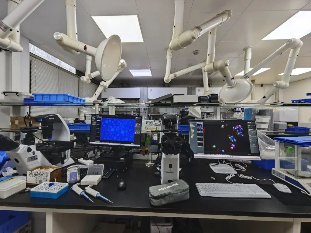
MSHOT MIX60-FL inverted fluorescence microscope with LED fluorescence illumination.
Consistent illumination settings
MIX60-FL provides independent illumination intensity memory for each channel.
When users switch between channels, they can return to previously selected light levels without repeatedly adjusting the illumination. This is useful when collecting images of different labels within the same sample.
The system also supports phase contrast, allowing researchers to examine morphology and fluorescent signals on one platform.
What matters for fluorescence quantification?
Fluorescence capability alone does not establish a quantitative imaging workflow.
Reliable comparisons also require control of:
Illumination intensity and exposure time
Camera gain and image acquisition settings
Background signal and chip autofluorescence
Photobleaching during repeated acquisition
Image processing and analysis methods
Chip material is particularly relevant. Background fluorescence from the substrate or surrounding materials can reduce the visibility of weak signals.
MIX60-FL is an option for laboratories that need fluorescence imaging alongside routine brightfield and phase contrast observation. The complete configuration should be selected around the fluorophores, signal levels, and measurement requirements.
4. Long-Term Live Cell Imaging in Microfluidic Chips: MCS31
Many microfluidic studies focus on biological changes that take hours or days.
Drug responses, cell growth, co-culture interactions, and organoid development require repeated observations. Manually returning to the microscope at every time point can be difficult, especially for overnight experiments.
Automating image collection
The MSHOT MCS31 Live Cell Imaging System supports scheduled imaging, multichannel acquisition, and time-lapse monitoring.
Its compact inverted design supports operation inside a compatible CO₂ incubator, helping researchers collect images while maintaining appropriate culture conditions.
For microfluidic use, the setup must also accommodate the chip, holder, tubing, and fluid delivery arrangement. These should be checked as a complete system before beginning a long experiment.
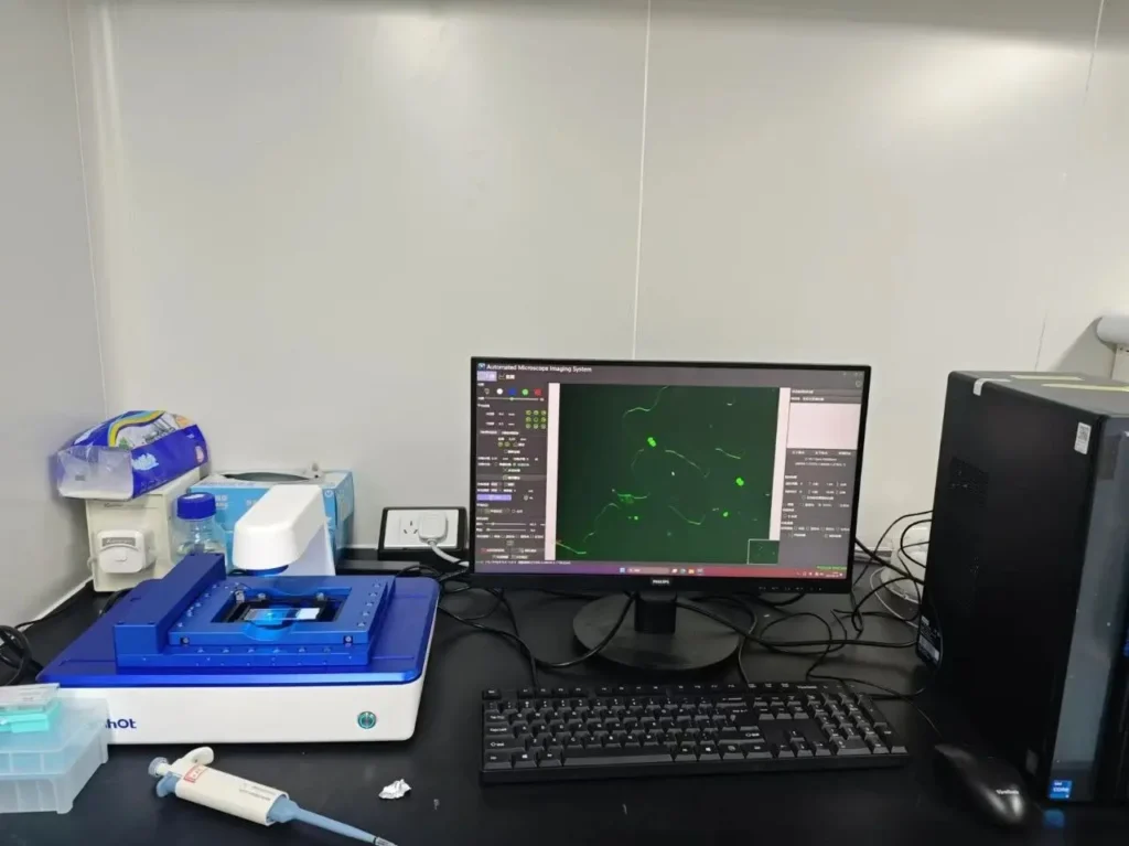
MSHOT MCS31 Live Cell Imaging System.
MCS31 supports Z-stack acquisition, with a minimum focus step of ≤1 μm and stacks of more than 50 image planes.
This allows samples to be recorded at different focal depths. Depending on sample transparency and optical compatibility, it can support the observation of cell aggregates and changes in organoid morphology.
The focus step describes the movement between image planes; it should not be interpreted as the system’s optical resolution.
Reducing repetitive analysis
MCS31 is worth considering for:
Overnight or multiday cell imaging
Monitoring cellular responses to drug exposure
Recording microfluidic co-culture experiments
Observing organoid-on-chip morphology over time
Performing plate-based preliminary studies
Its main advantage is maintaining a consistent image acquisition schedule while reducing repeated manual work.
5. Working Distance, Chip Thickness and Camera Speed: What to Check Before Buying
Selecting a microscope for microfluidics involves more than choosing magnification or fluorescence channels. Several practical checks determine whether the system will work with the actual experiment.
Chip bottom thickness and objective compatibility
For inverted imaging, the objective must focus through the bottom of the chip.
Check the bottom material and thickness, the distance from the external surface to the sample, and the objective’s specified working distance and correction requirements.
An objective designed for one bottom thickness may not provide the same image quality through a different chip construction. Higher magnification does not automatically solve poor optical compatibility.
Objective and condenser working distances
Objective working distance and condenser working distance describe different clearances.
The objective working distance affects access to the imaging plane from below. The condenser working distance affects the transmitted-light arrangement above the sample.
For example, the MIX60 platform’s 72 mm condenser working distance provides clearance above the sample. It does not mean that the objective can image through a 72 mm chip bottom.
Space for tubing and chip holders
Confirm that the complete assembly fits on the stage.
Consider the holder footprint, observation window position, connector height, tubing bend radius, and strain on the chip during stage movement.
A fixture that fits physically may still prevent the objective from reaching the required observation area.
Camera speed and sensitivity
Fast flow events and slow biological changes place different demands on the camera.
Droplet generation may require rapid acquisition and short exposures. Weak fluorescence may require greater sensitivity and longer exposures.
Choose the camera and acquisition settings around the process you need to record.
Culture conditions and experiment duration
For long-term cell experiments, consider temperature, CO₂, evaporation, focus stability, and fluid delivery continuity.
Plan the imaging interval, number of positions, fluorescence channels, and Z-planes together. Adding more images to each cycle increases acquisition time and may increase light exposure.
6. Frequently Asked Questions About Microscopes for Microfluidics
Do microfluidic experiments always require an inverted microscope?
No. An inverted microscope is often convenient for transparent chips with fluid connections above the sample. An upright reflected-light microscope may be more suitable for inspecting opaque molds, electrodes, and accessible surface structures.
Is fluorescence necessary for observing microfluidic flow?
Not always. Brightfield imaging may be sufficient for visible droplets, particles, or channel features. Fluorescence is useful when the experiment requires labeled targets or fluorescence-based detection.
Can one microscope provide both phase contrast and fluorescence?
Yes. A suitably configured system such as MIX60-FL supports both methods. Researchers can examine sample morphology and fluorescent labels on the same platform, with settings adjusted for each imaging mode.
Does a long-working-distance objective work with every chip?
No. Compatibility also depends on bottom thickness, optical correction, sample depth, chip material, and the required image quality. Check the actual chip construction against the objective specifications.
What is the difference between time-lapse imaging and high-speed imaging?
Time-lapse imaging records changes at selected intervals over an extended period, such as cell growth over 48 hours. High-speed imaging records rapid events, such as droplet formation, using a sufficiently high frame rate.
Can automated cell analysis be used with microfluidic chips?
It may be suitable, but the analysis should be validated on representative images. Chip structures and background features can influence how software identifies and measures cells.
Choosing the Right Microfluidics Imaging System
For fabrication inspection, MJ31 supports reflected-light observation of suitable molds, electrodes, and surface features.
For routine transparent sample observation, MIX60 provides brightfield and phase contrast imaging. When fluorescent labels are central to the experiment, MIX60-FL adds multichannel fluorescence capability.
For studies that require repeated imaging over hours or days, MCS31 provides scheduled acquisition and analysis tools for compatible samples and assays.
The most useful starting point is a clear description of the experiment: what the chip contains, how it is constructed, what must be measured, and how quickly or how long the process needs to be recorded.