Cancer


Cancer generally arises from genetic mutations or abnormal gene expression in cells, leading to abnormal cell differentiation and proliferation, uncontrolled growth. It can invade adjacent parts of the body and spread to other organs.
Pet Hospital front

Microscopic Imaging Applications in Cancer Research


Based on the changes of cancer cells at the molecular expression level and cellular tissue level, various microscopic imaging techniques are employed to screen and identify cancer cells, such as: ① Pathological Sections; ② Immunohistochemistry (IHC); ③ Fluorescence In Situ Hybridization (FISH); ④ Circulating Tumor Cell Detection (CTC); ⑤ Tumor Cell Culture and Research.

Challenges for Microscopic Imaging in Cancer Research


There are two types of imaging in cancer research: brightfield imaging and fluorescence imaging. Brightfield imaging has low equipment requirements, but the full-slide interpretation of pathological sections involves heavy workload and low efficiency. A slide scanning system with electric scanning and image stitching software can realize efficient global imaging and local magnification, improving diagnostic accuracy and efficiency.
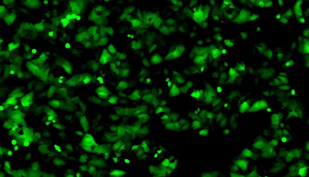
Multicolor fluorescence imaging features high sensitivity and specificity, but suffers from weak signals. Obtaining clear multi-channel signals requires optimization of excitation light, filters, imaging equipment and image processing.
For cell culture, there are also difficulties in long-term imaging monitoring, while reducing contamination and avoiding impacts on cell growth.

Solutions for Microscopic Imaging in Cancer Research

  • 1. All-Round Equipment: Sunny MDS4+MSX2/MS60 Digital Slide Scanning System, integrating slide scanning function and research-grade fluorescence microscope, suitable for pathological section, CTC, FISH, IHC and other detection scenarios.
  • 2. Specialized Equipment for FISH, CTC and IHC: MF43-N/MF53-N+MS23/MSX11, equipped with FISH dedicated software, supports multi-color fluorescence channels, realizes high-sensitivity imaging and intelligent image optimization.
  • 3. Observation of Cancer Cell Culture: MCS11/MCS21/MCS22 Live Cell Imager with computer, can be used in incubators with wireless connection and remote management, reduces contamination risk, and integrates multiple imaging modes to meet diverse demands of cell culture observation.

Product Recommendations