Home » Microscopy Applications for NMPA Drug Safety Evaluation Labs
Guardian of Drug Safety — Exploring Microscopy Applications in NMPA Drug Safety Evaluation Laboratories
Against the backdrop of growing public demand for health, drug safety has become the focus of global attention. In the chain of drug research and development and production, the Drug Safety Evaluation Laboratories of the NMPA act as a guardian, shouldering extremely important responsibilities.
As a core instrument in the laboratory, microscopes play a key role in all aspects of drug safety evaluation with their powerful observation capabilities, providing a solid guarantee for assessing the safety and efficacy of drugs.
Overview of Microscopy Applications in Drug Safety Evaluation
Using pathological techniques such as HE staining and Masson staining, high-resolution microscopes are employed to observe cellular lesions (e.g., inflammation or necrosis) in experimental animal tissue sections, providing visual evidence for the evaluation of drug toxicity.
Challenges of Microscopic Imaging in Drug Testing
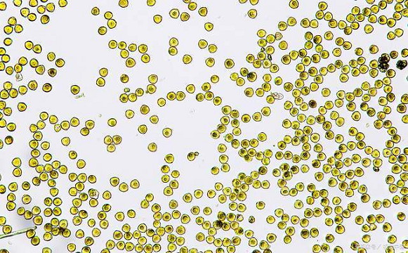
In drug safety evaluation, microscopic imaging technology faces several challenges. Firstly, the detection of microbial limits in pharmaceuticals and the observation of toxicopathological sections mostly rely on brightfield imaging. Given the small size and low quantity of microbial particles, as well as the typically tiny lesion areas in tissue sections, high-resolution and high-contrast imaging systems are required. Secondly, in neurotoxicology testing, it is necessary to capture subtle structural changes such as synapses and axons, which imposes high demands on the resolution and signal-to-noise ratio of the microscopic imaging system.
Drug Microbial Limit Test / Toxicopathological Section Observation
Brightfield Observation Solution
Recommended Configuration: Upright biological microscopes ML45 / ML51‑N / ML31 + microscope cameras MDX10 / MD60 Suitable for brightfield observation of Staphylococcus, Vibrio cholerae, Candida and other microorganisms.
Intelligent Digital Biological Microscope ML45
- Magnification: 40X–1000X
- Coded objective turret
- Brightness memory function
- LED digital display showing current working status
- ECO auto power-off (lights off when user leaves)
- Upgradable for phase contrast, darkfield, simple polarized light and fluorescence observation
Microscope Camera MDX10
- 20-megapixel high resolution
- 1‑inch large format sensor
- 22 fps at full resolution
- Balances high-definition imaging and high speed


