
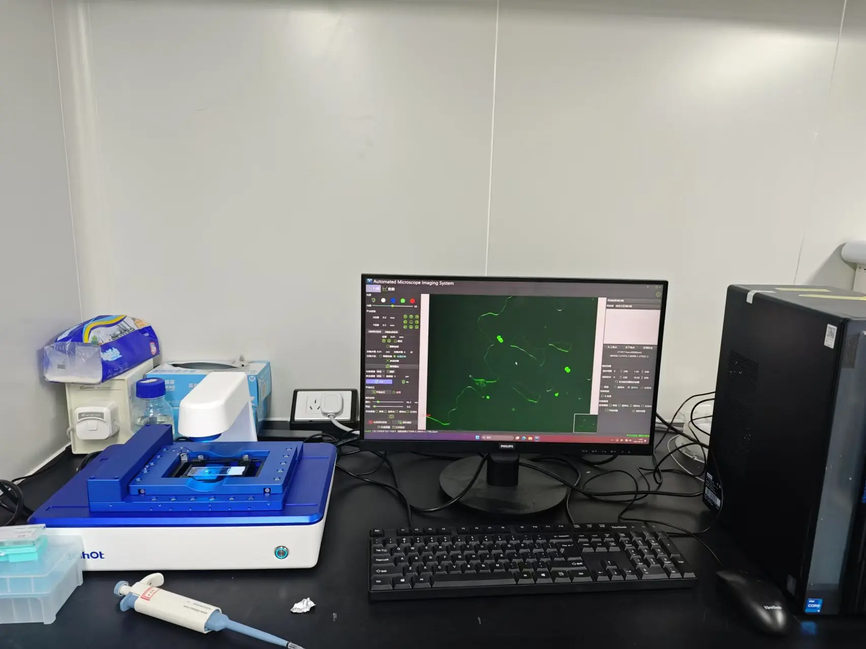
Are You Recording Cell Behavior—or Experimental Artifacts?
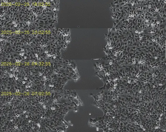
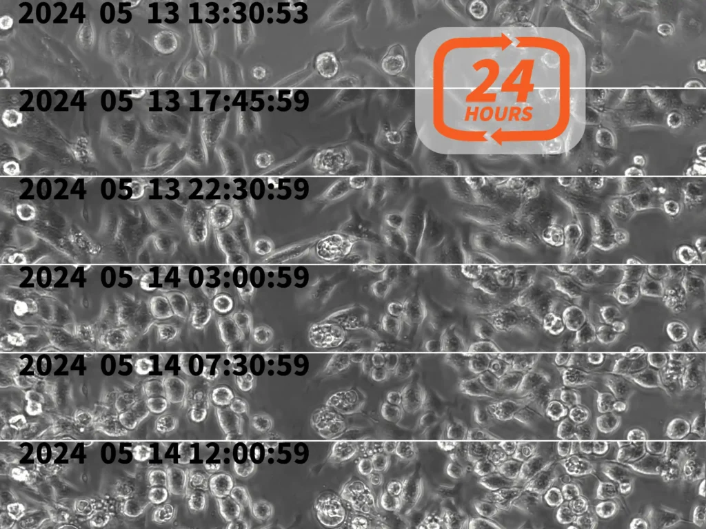
In long-term live cell imaging, changes in morphology, migration or proliferation do not always originate from the cells themselves.
Temperature fluctuations, repeated sample transfer, focus drift, inconsistent imaging positions and excessive fluorescence exposure can all affect the results.
To generate reliable time-lapse data, several technical variables must be controlled throughout the experiment.
1. Environmental Stability
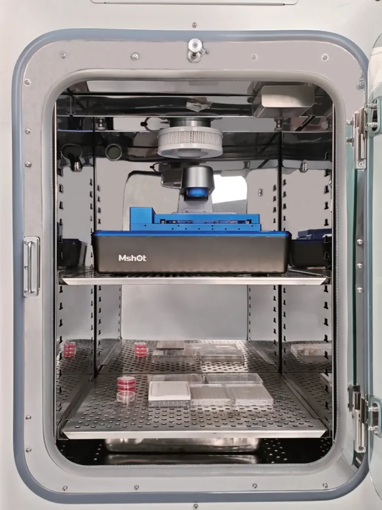
Moving culture vessels repeatedly between an incubator and a conventional microscope may disturb temperature and gas equilibrium. Frequent handling also increases contamination risk.
These environmental changes can influence cell adhesion, morphology, proliferation and migration. In experiments lasting several hours or days, even minor disturbances may accumulate and reduce comparability between time points.
Imaging should therefore be performed with minimal interruption to the established culture environment.
2. Positioning and Focus Repeatability
Time-lapse analysis requires the same sample region and focal plane to be captured repeatedly.
In multiwell experiments, manual positioning can introduce differences in the field of view. Focus drift may also alter cell boundaries, image contrast and measured confluence, making it difficult to separate biological changes from imaging variation.
Motorized XYZ positioning, predefined coordinates and autofocus improve acquisition repeatability, particularly when multiple wells and imaging positions are recorded in each cycle.
3. Representative Sample Coverage
Cells are rarely distributed uniformly across an entire well. A single field of view may therefore provide an incomplete or biased representation of the sample.
Multipoint imaging can collect data from several predefined positions, while selected-area scanning and image stitching can extend observation beyond one microscope field.
This is particularly useful for:
- Cell confluence measurements
- Scratch-wound assays
- Cell migration studies
- Multiwell treatment comparisons
- Organoid growth monitoring
4. Temporal Resolution and Fluorescence Exposure
The acquisition interval must match the speed of the biological process.
Short intervals provide higher temporal resolution but generate more data and increase light exposure. In fluorescence experiments, excessive illumination may cause photobleaching, signal reduction or phototoxic effects.
Phase contrast can be used for routine label-free monitoring, while fluorescence channels can be added only when labeled cells or structures need to be identified.
Exposure time, illumination intensity and acquisition frequency should be optimized before long-term recording begins.
How MCS31 Addresses These Requirements
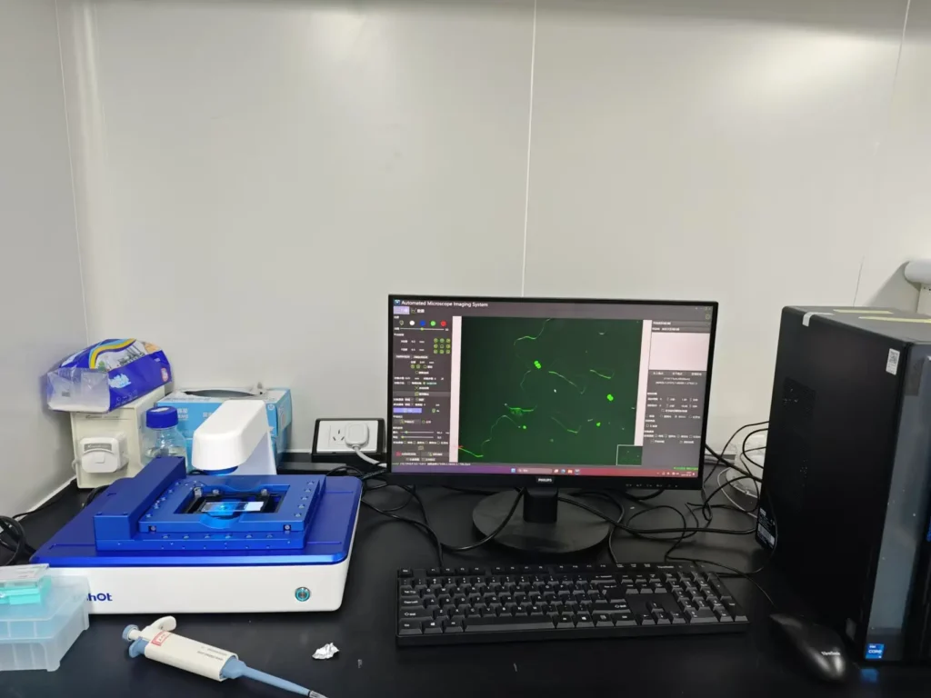
he MSHOT MCS31 Live Cell Scanning and Analysis System is designed to operate inside a compatible cell culture incubator. Cells can remain under established culture conditions while the system performs automated time-lapse imaging.
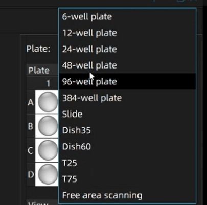
The MCS31 supports culture flasks, culture dishes and multiwell plates from 6 to 384 wells, allowing it to accommodate both routine cell culture and multiwell comparative experiments.
Its imaging capabilities include:
- Motorized XYZ positioning
- Autofocus
- Multipoint acquisition
- Selected-area scanning and image stitching
- Motorized dual-objective switching
- Phase-contrast imaging
- Blue, green and UV fluorescence channels
Researchers can define imaging wells, positions, channels and acquisition intervals according to the experimental design. The system then returns to the selected positions automatically during each acquisition cycle.
The integrated software supports:
- Cell counting
- Cell confluence analysis
- Scratch-wound assay analysis
- Cell viability analysis
- Time-lapse imaging and video generation
- Field-of-view control
- Email notifications
Optional AI analysis functions can also be expanded for specific workflows.
From Image Acquisition to Repeatable Measurement
Reliable live cell imaging requires environmental stability, repeatable positioning, controlled illumination and an analysis method matched to the experimental endpoint.
By combining incubator-compatible operation, automated scanning, phase-contrast and fluorescence imaging, autofocus and integrated analysis, the MCS31 reduces sample handling and improves the consistency of long-term observation.
It is suitable for cell growth monitoring, migration assays, fluorescent transfection studies, organoid culture and drug-response experiments.