Description
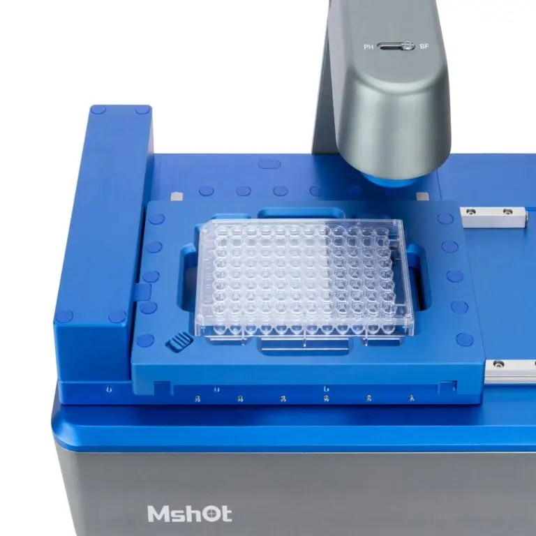
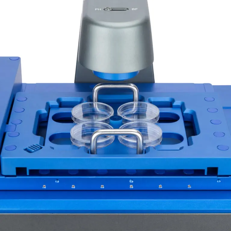
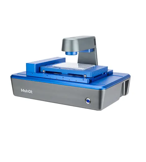
The Live Cell Imaging System MCS31 is an excellent companion for cell culture. It can be used inside an incubator and supports phase contrast and fluorescence observation of various vessels, from culture flasks and dishes to 6-well and 384-well plates, enabling long-term, non-invasive monitoring of living cells. As a trusted name in microscopy, MSHOT designs the Live Cell Imaging System MCS31 to meet the practical needs of modern laboratories, combining dependable optics with straightforward operation.
Overview
MSHOT designs cell-friendly imaging systems that fit directly into the researcher’s workflow. The Live Cell Imaging System MCS31 is built around a well-engineered optical platform that delivers consistent, high-quality live cell imaging system results in routine and research settings. Every component, from the objective system to the illumination source and the mechanical stage, has been selected and calibrated to provide stable, repeatable imaging session after session.
Whether you are a researcher running long experiments, a laboratory technician processing many samples, or an educator demonstrating principles to students, the Live Cell Imaging System MCS31 is designed to make live cell imaging system work more efficient and more reliable. Its thoughtful layout and clear controls reduce setup time, while robust construction ensures it stays accurate even under continuous use.
Key Features
- Works inside standard incubators for stable, long-term time-lapse imaging
- Dual phase-contrast and fluorescence observation modes
- Supports culture flasks, dishes and multi-well plates (6-well to 384-well)
- Compact, easy-to-use design with repeatable positioning
- Automated image capture for continuous live cell experiments
These features work together to simplify your live cell imaging system workflow. The optical system delivers sharp, well-corrected images across the field of view, while the illumination and observation modes give you the flexibility to examine different sample types with confidence. The result is an instrument that performs consistently, whether you are looking at prepared slides, living cultures or industrial specimens.
Maintenance is equally straightforward. The durable construction and accessible design make cleaning and routine care simple, so the Live Cell Imaging System MCS31 remains dependable throughout its service life with minimal downtime.
Applications
Thanks to its balanced design, the Live Cell Imaging System MCS31 is well suited to a broad range of live cell imaging system tasks:
- Long-term cell proliferation and migration studies
- Live cell morphology tracking over hours or days
- Label-free phase-contrast monitoring of cell cultures
- Fluorescence-based reporter assays in living cells
Across these applications, users benefit from stable imaging, consistent color rendition and a workflow that minimizes adjustment time. The Live Cell Imaging System MCS31 adapts readily to different specimen preparation methods and observation requirements, making it a versatile addition to any laboratory.
Build Quality and Ergonomics
Built with precision components and a solid frame, the Live Cell Imaging System MCS31 offers the stability needed for critical observation. Its ergonomic layout places controls within easy reach, reduces operator fatigue and allows comfortable use over extended sessions. This attention to detail helps you obtain more accurate results with less effort.
Specifications
The MCS31 integrates a precision motorized stage and software-driven acquisition, allowing multiple culture vessels to be imaged sequentially without disturbing the incubation environment. For complete technical specifications, including optical configuration, illumination options and available accessories, please refer to the product datasheet or contact our team.
For laboratories studying dynamic biological processes, the MCS31 supports automated time-lapse capture that runs unattended while samples remain safely in the incubator. The system records changes in cell morphology, confluence and fluorescent signals, giving you a complete picture of culture behavior over time.
Why Choose MSHOT
MSHOT is a professional microscope manufacturer committed to combining optical excellence with responsive support. Every live cell imaging system solution is backed by clear documentation, firmware updates and practical application guidance. Our team helps you select the right configuration, supports installation and remains available for questions as your needs evolve.
Contact our team today to discuss your requirements, request a quotation, or arrange a demonstration of the Live Cell Imaging System MCS31 for your laboratory.