Live Cell Imaging System MCS31

The Live Cell Imaging System MCS31, an excellent cell culture companion, works in incubators. It supports phase contrast and fluorescence observation of various vessels (from culture flasks, dishes to 6-well to 384-well plates), and enables scanning/stitching of selected regions for large-field, long-term multi-well plate recording. This boosts efficiency in applications like comparative experiments and reduces contamination-related failure risks.
Share:

Get Exclusive Offers and Updates!

    Description

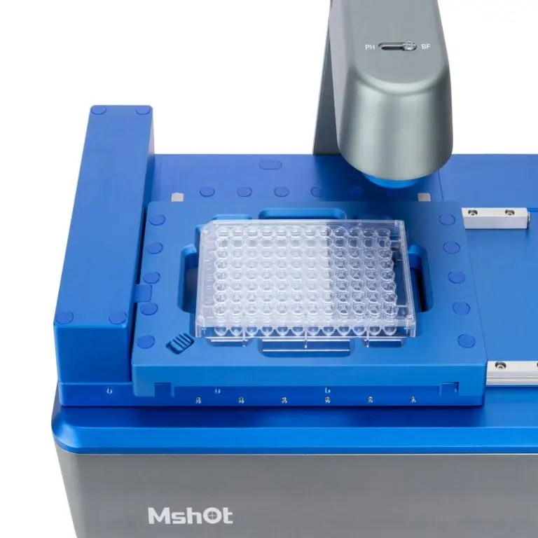
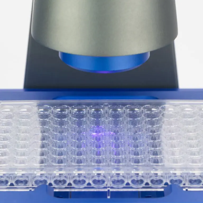
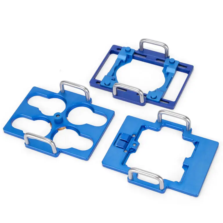
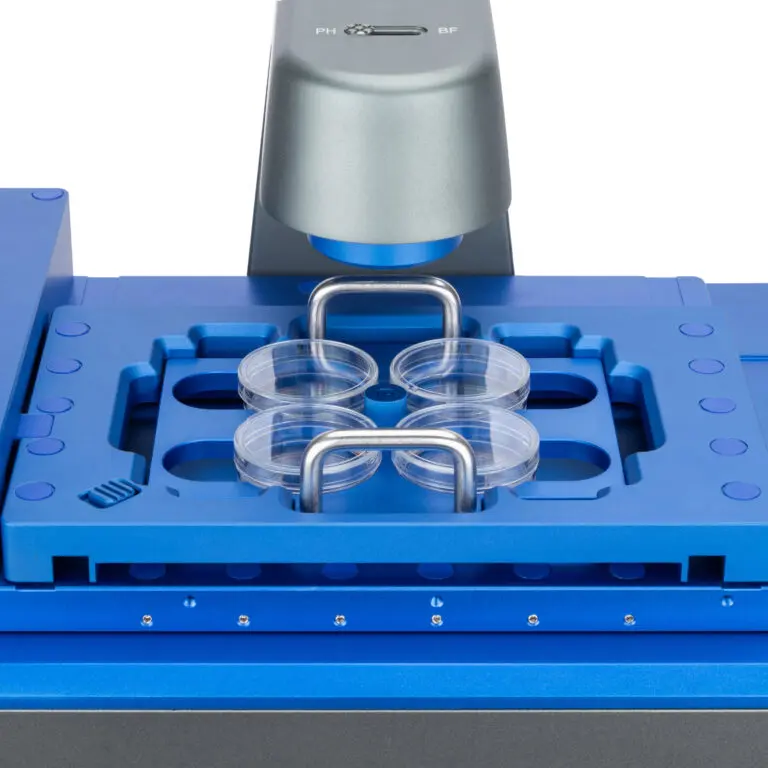
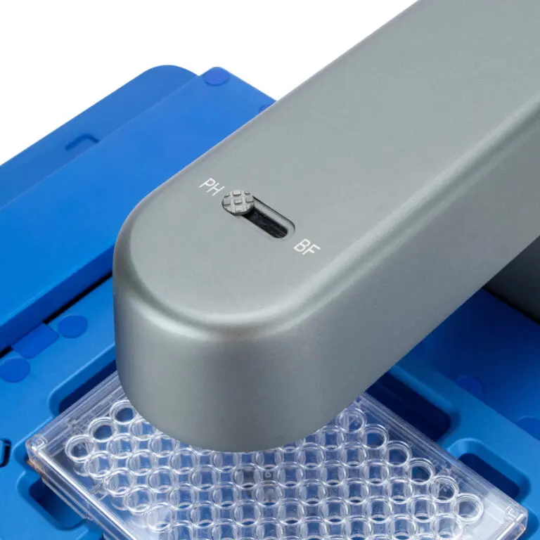
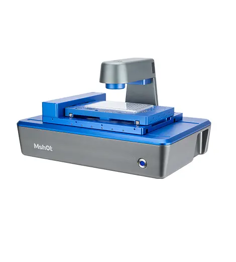
    The Live Cell Imaging System MCS31 is an excellent companion for cell culture. It can be used inside an incubator and supports phase contrast and fluorescence observation of various culture flasks, petri dishes, as well as 6-well to 384-well plates. It enables scanning and stitching of selected regions, allowing for large-field-of-view, long-term observation and recording of multi-well plates. This significantly improves experimental efficiency in application scenarios such as comparative experiments and reduces the risk of experimental failure caused by contamination and other factors.

    Feature

    Long-term imaging in incubators

    Compatibility with multi-well plates

    Motorized switching between dual objectives

    Detail

    Specifications

    Item Specifications
    Brightfield Observation Transmitted phase contrast illumination, working distance 55mm
    50,000 – hour long lifespan, 625nm low – phototoxicity LED light source
    Fluorescence Observation
    (Fluorescence Wavelengths Customizable)
    ·Blue (B): 472 – 495nm
    ·Green (G): 543 – 560nm
    ·Ultraviolet (UV): 393 – 416nm
    Objective Lenses ·Select any two from 4×/10×/20× objective lenses; Motorized objective lens switching
    Stage Motorized XY moving platform with a stroke of 115*77mm; Repeat positioning accuracy ≤ 3μm
    Z – axis Motorized auto – focus mechanism
    Camera High – speed and high – sensitivity camera, 500W 2/3 inch, 40fps
    Software Supports remote control, including light control, camera control, photo taking, video recording, time – lapse photography
    Analysis Functions Cell counting, cell confluency, scratch assay, transfection efficiency, viability analysis
    Scanning Functions ·Multi – well imaging: Custom – set XY array for multi – point photography, brightfield and fluorescence multi – channel overlay
    ·Region scanning: Set rectangular regions for full – scan stitching, brightfield and fluorescence multi – channel overlay
    Data Transmission/Power Supply USB data cable + DC power cable
    Working Environment 5 – 40°C, 20 – 95% RH
    Dimensions 252mm × 377mm × 208mm

    Resources

    https://www.m-shot.com/unlocking-the-mysteries-of-the-brain-how-the-mshot-mcs31-breaks-through-the-three-major-bottlenecks-in-microglial-cell-research/