Pain is a shared “nightmare” for patients with vascular diseases, yet the molecular mechanism behind it has long remained an unsolved mystery in the medical field.
Recently, a research team led by Professor Liu from the School of Pharmacy, Nantong University, in collaboration with multiple domestic institutions, published a landmark study in the international journalAdvanced Science(Impact Factor: 14.3). This study reveals, for the first time, a novel mechanism by which vascular endothelial cells regulate vascular pain through the ET-1/ETAR signaling pathway.
The study leveraged advanced equipment such as Mshot’s inverted fluorescence microscope MIX60-FL to successfully decipher the unique mechanism of vascular pain, providing a new target for clinical pain treatment.
Mshot offered reliable technical support for this major discovery, once again demonstrating the value of domestic high-end microscopes in cutting-edge life science research.
Research Highlights
Innovative Model Defines a New Type of Pain
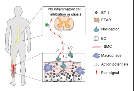
For the first time, the team successfully established a vascular pain animal model by ligating the superficial blood vessels in the hindlimbs of rats. The study found that vascular pain only presents as mechanical hyperalgesia, with no thermal or cold hyperalgesia, nor immune cell infiltration or glial cell activation—confirming it as a new type of pain independent of neuropathic pain and inflammatory pain.
Mechanism Breakthrough: Key Pathway Identified
Using optogenetics, chemogenetics, and transgenic technologies, the team found that ET-1 (endothelin-1) derived from vascular endothelial cells activates the ETAR receptor on nociceptive sensory neurons, forming an “endothelium-nerve-spinal cord” regulatory loop that drives the development of vascular pain. This discovery provides a new target for clinical treatment.
Clinical Translation: Benefiting Patients
The study further verified that oral administration of bosentan, an ETAR antagonist, can significantly alleviate tourniquet-induced vascular pain in clinical patients, bringing a novel strategy for pain treatment.
Mshot MIX60-FL: The Hero Behind Clear Microscopic Imaging
The groundbreaking achievements of the Nantong University team would not have been possible without the support ofMshot’s intelligent LED inverted fluorescence microscope MIX60-FL. With the following advantages, this equipment has become a “golden partner” in life science research:
LED Multi-Color Fluorescence Imaging
It features a modular 3-color fluorescence system (upgradeable to 4 colors) paired with a digital-display LED light source, which has independent light intensity memory. This ensures high sensitivity and repeatability of fluorescence signals, helping the team clearly capture the dynamic expression of ET-1 in endothelial cells. With an ultra-long lifespan of 50,000 hours, it supports instant on/off operation—simple and easy to use.
Intelligent Interaction: Efficient and Convenient
The built-in OLED digital display shows objective magnification and light intensity in real time. The voice broadcast function enhances operational efficiency, allowing researchers to focus on scientific questions rather than equipment adjustment.
Semi-Phase Contrast Fluorescence Objectives: Every Detail Revealed
The high-performance objectives meet the observation needs of both fluorescence and transparent samples with phase contrast, providing high-resolution image support for vascular-nerve interaction research.